Photo courtesy of Fujifilm
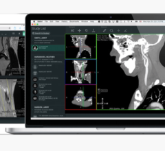
December 3, 2014 — Fujifilm will showcase the ability of Synapse 3-D and discuss how radiologists use 3-D models to shape the future of patient care at the 100th annual meeting of the Radiological Society of North America (RSNA) in Chicago, Ill.
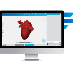
"Radiologists are putting patients at the center of care with the use of 3-D modeling in surgical treatment planning. By leveraging image overlay tools available on Synapse 3D, radiologists can use volumetric imaging to generate 3-D models and quantitative analysis of organs and other parts of the anatomy," said Jim Morgan, vice president of Medical Informatics at Fujifilm Medical Systems Inc. "Referring physicians and radiologists can then leverage the images and data to develop treatment plans, predict and prepare for complications during procedures, as well as educate patients about their conditions prior to treatment."
At Keck Hospital, USC Norris Comprehensive Cancer Center and Hospital in Los Angeles, radiologists scan an estimated 400 patients annually who present for a consultation for renal cancer surgery, and around 300 of those patients eventually undergo surgery. Radiologists and surgeons there use Synapse 3D to develop virtual 3-D models for the evaluation of renal masses and to develop treatment plans.
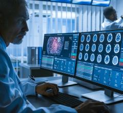
The hospital's Abdominal Imaging Section Chief and Medical Director of Imaging, Vinay A. Duddalwar, M.D., FRCR, describes a recent case in which he developed a 3D model for surgery on a patient with a renal mass. The patient presented with pain in the abdomen and with bleeding in his urine. After undergoing a single-phase CT scan at another center, he was referred to doctors at the USC Norris Comprehensive Cancer Center and Hospital where he underwent a standard of care multiphase-CT scan. Dr. Duddalwar used Synapse 3D to evaluate the study.
"We have standard protocols in Synapse 3D that we use to enhance the visualization of the mass and extract information that the urologist then uses. In this particular case, we looked at the prior CT scan, but evaluated the multiphase-CT scan of the tumor on Synapse 3D, using all of the phases in Synapse," said Duddalwar, M.D.
He created the virtual 3-D model by registering all four-volume datasets. "Synapse 3D allows you to overlay and fuse the images from phases one, two, three, and four together. It allows the user to subtract the blood vessels or subtract the collecting system from the kidney so we can see only the functioning parenchyma or only the blood vessels," Duddalwar explains. "It allows us to configure different visualization streams so you can see everything, extract features or superimpose another, making the tumor transparent."
"Synapse 3D gave us the diagnostic confidence that we needed to determine that it was a low-grade tumor. The reasons were, one, it didn't enhance very much; two, it was fairly uniform; and three, its shape was small and round. The virtual 3D models helped in conveying this information to the surgeon," said Duddalwar. "Synapse 3D helped increase our diagnostic confidence in determining this was a low-grade tumor and in explaining our recommendation to the surgeon and the patient."
Synapse 3D offers more than 30 clinical functions to choose from, with many functions utilizing Fujifilm's Image Intelligence capability. It is fully integrated with Synapse PACS and Synapse Cardiovascular, and creates a centralized enterprise-shared 3-D solution. The Synapse 3D snapshot workflow creates a unique closed loop workflow from modality labs, to Radiologists and Cardiologists, to Surgery with no redundant rendering work required along the way, creating faster workflows.
For more information: www.fujimed.com


 April 18, 2025


