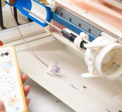
Dr. Nicholson says using the Celero device means faster procedures and more
With more than 550,000 core needle biopsy procedures performed annually in the U.S., there is a real need to match the right biopsy device – one that is both effective and efficient – to a given procedure.
For the radiologist and surgeon, the right device is easy to use, can be used with a broad range of cases and gathers plenty of tissue without complications. And for the patient, the right device makes the procedure faster, less painful and less traumatic. This is particularly important since 80 percent of all biopsies are benign and can help women avoid an invasive open surgical procedure if correctly diagnosed with percutaneous biopsy.
A new 12-gauge handheld, the Celero vacuum-assisted spring-loaded core device was recently introduced by Suros Surgical Systems Inc., a Hologic company, for use in the U.S. ultrasound breast biopsy market. The device has the ability to fire the inner cannula both inside and outside the breast, a feature women’s health doctors have been looking for and other companies have tried unsuccessfully to develop. The larger core samples from the Celero handpiece allow these biopsy procedures to be faster and less traumatic for the patient due to fewer needle insertions. Larger and more contiguous samples also increase the opportunity for the pathologist to make an accurate diagnosis.
Biopsying Hard-to-Reach Breast Lesions
Mary Nicholson, M.D., a fellowship-trained breast radiologist at the Women’s Diagnostic Center in Munster, IN, was one of the first radiologists to evaluate the Celero device. “One of my first cases was an abnormally enlarged, low-lying axillary lymph node deep within the tissue,” said Dr. Nicholson. “In four passes, I had gathered plenty of tissue and was done, without complications.”
Dr. Nicholson indicated that the Celero device is particularly good for use with challenging lesions, such as those in the axilla, close to the chest wall, near implants or behind the nipple. Lesions located in these areas can make even the most confident physician hesitant to perform a biopsy.
In the post-fired position, the Celero needle is easily advanced through the most dense breast tissue. If physicians choose to enter the breast before firing the device, they can place the trocar tip directly adjacent to the lesion and then fire the device. The trocar tip allows the Celero needle to fire in a direct, straight line, without diving away in the tissue acquisition process like some other spring-loaded core devices.
The Celero device is designed to be an easy one-step setup, and because it is fully disposable, cleanup is just as easy. The highly echogenic needle composition makes it easy to see under ultrasound and clearly verifies accuracy of biopsy site targeting. The Celero device was designed to meet the needs of physicians who currently use spring-loaded devices under ultrasound guidance. Held in place by a 20inHg
vacuum, the captured tissue in the Celero aperture (sample notch) isconsistently two to three times the sample amount of traditional spring-loaded core devices.
A Clinically Better Solution
According to Dr. Nicholson, “The lightweight Celero device is easy to maneuver, and the ergonomically designed handpiece has the collection and firing buttons positioned in such a way that they can be easily pressed, all while maintaining position within the tissue. I can count on the samples coming back from pathology with a definitive diagnosis with the Celero device. The larger core also means I can enter the breast with the device fewer times, resulting in a faster biopsy procedure and more compassionate patient care.”
Because the inner cannula can be fired before entering the breast, physicians can gently move to the lesion and precisely place the sample notch where desired. Once in or near the lesion, the physician can confirm placement of the aperture with ultrasound imaging prior to firing the outer cannula. The sample is then acquired safely, without harming the chest wall, nipple or any other sensitive area. “I believe the device’s option of firing outside the breast works remarkably well for lesions in these challenging areas, as well as for typical ultrasound breast biopsy procedures,” she said.
For physicians who prefer to use an introducer sheath, the Celero system comes with its own introducer, which snaps onto the needle and is inserted into the breast with the device. The introducer can then be unsnapped, leaving it in the needle track and making it convenient for easy insertion and deployment of the CeleroMark, a new end-deploy titanium marker for use with the Celero handpiece.
The Celero device is the ideal solution for dense breast tissue, which can offer resistance to needle penetration, according to Dr. Nicholson. In using it on a patient whose breast tissue was mammographically dense and who was suspected of having a fibroadenoma in her breast, she inserted the Celero needle into the breast tissue, easily navigated and traversed through the dense breast tissue, and acquired a sample with little to no resistance. “In total, four samples were obtained easily from tissue that typically presents difficulties with traditional spring-loaded core biopsy devices,” she noted.
Dr. Nicholson emphasized that the Celero breast biopsy device has exceeded her expectations. “This device’s highly echogenic aperture can be precisely placed for target verification prior to tissue acquisition. Its sharp tip is capable of penetrating all mammographic compositions of breast tissue. The Celero needle offers less resistance in all types of breast tissue and performs better than other spring-loaded biopsy devices. And the lightweight design of the handpiece and its trocar needle tip provides smooth penetration into lesions, reducing chances of deflection.”
For the breast ultrasound market, the Suros Celero disposable biopsy device offers a superior clinical solution.


 February 18, 2026