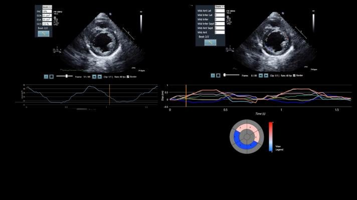
April 9, 2019 — DiA Imaging Analysis announced the launch of LVivo SAX, a cardiac analysis tool that helps clinicians quickly and accurately interpret ultrasound images and assess heart functionality among patients suspected of suffering from acute coronary syndrome (ACS).
LVivo SAX uses artificial intelligence (AI) to analyze segmental left ventricle wall motion using the parasternal short axis view. This is a common cardiac view used in point-of-care ultrasound settings, as it provides views of cardiac tissue supplied by all three major coronary vessels and is relatively easy to acquire without manipulating the patient’s posture. The LVivo SAX tool is designed to provide medical clinicians with varying levels of ultrasound analysis or cardiological experience the ability to automatically measure, track and evaluate cardio functions, and to detect abnormalities that reduce variability and increase efficiency.
Emergency room and point-of-care clinicians typically order an electrocardiogram (ECG), a blood test and an ultrasound when a patient presents with ACS symptoms. The ultrasound images, which are vitally important given that ECG and blood tests are not always conclusive, are typically viewed visually. The results are often dependent on the clinician’s level of training and experience. As an integrated part of an ultrasound and information technology (IT) system, LVivo SAX leverages AI to bring objectivity to the process to provide an automated and objective assessment of left ventricular function and segmental function.
“LVivo SAX has the potential to change the way emergency department clinicians manage patients with suspected ACS,” said Chris Moore, M.D., associate professor of emergency medicine at Yale University School of Medicine. Moore will be conducting a post-market clinical study of LVivo SAX within Yale’s emergency room environment.
“Today, the accurate assessment of ultrasound images often depends on the expertise of the interpreter. By delivering reliable and reproducible information related to wall motion abnormalities, LVivo SAX helps doctors arrive at the correct diagnosis and guide the proper care of the patient. We are pleased to take part in DiA’s launch of LVivo SAX and look forward to trialing the tool in our emergency department,” Moore said.
DiA will demonstrate LVivo SAX, as well as its extended LVivo Cardiac Toolbox, at the American Institute of Ultrasound in Medicine (AIUM) 2019 conference, April 6-10 in Orlando, Fla.
For more information: www.dia-analysis.com

 October 07, 2026