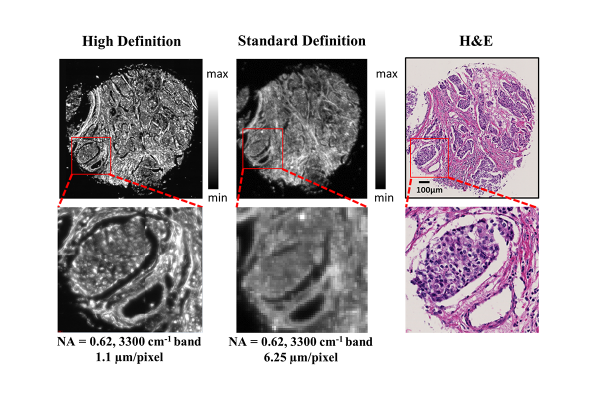
Comparisons of high definition and standard definition infrared imaging for digital histopathology. Image courtesy of the Beckman Institute
April 14, 2021 — Detecting and analyzing breast cancer goes beyond the initial discovery of the cancer itself. If a patient has a tumor removed and it needs to be analyzed to determine further treatment, it might be OK for the results to take 24 hours. But if the patient is still on the operating table and clinicians are waiting to make sure no cancer cells are present along the edges of the removed tumor, results need to be nearly immediate.
A paper titled, "Breast cancer histopathology using infrared spectroscopic imaging: The impact of instrumental configurations," was published in Clinical Spectroscopy, an extension of previous work by Beckman Institute Postdoctoral Fellow Shachi Mittal and Rohit Bhargava, bioengineering professor and director of the Cancer Center at Illinois. Two alumni of the Beckman Postdoctoral Fellows program, Michael Walsh and Tomasz Wrobel, are co-authors. It seeks to advance breast cancer detection methodologies when using spectroscopic imaging.
They conducted the research at the Beckman Institute for Advanced Science and Technology at the University of Illinois Urbana-Champaign.
Because time is of the essence in breast cancer detection, the research identifies how clinicians can choose the right detection methods and tools during the correct scenario to still drive accurate results.
"The goal of the study was to give people a roadmap of how to plan and design an infrared imaging-based study for clinical work like digital histopathology," Mittal said. "In one scenario, you determine what instrument configuration will be better, and then you determine what types of methodologies you can use to develop accurate models with that instrument."
The team set out to examine the tradeoffs between using a standard Fourier transform infrared image or a high-definition Fourier transform infrared image.
"We wanted to see how the different types of instruments, especially different resolutions and how it impacts the capability of that data set to be used for different diagnostic purposes," Mittal said.
Instead of determining that one method is always superior, the researchers uncovered that the answer is much more complex. While high-definition imaging may seem like the best option regardless of the circumstances, sometimes standard definition infrared spectroscopy suffices in accuracy in instances when clinicians need a speedy result, for example.
"As technology expands and provides more capabilities with new features, it becomes more difficult to choose the optimal technology from the many options available," Bhargava said. "This study provides a nice comparison and guidelines to design a more useful and practical technology."
For Mittal, the research is an important step towards better dissecting, understanding, and curing cancer.
"Cancer is something we understand very little about," she said. "It's not only about the journey of battling the disease, but also the mental burden. Patients oftentimes don't understand, and they recognize the doctor may not fully understand either."
Mittal began her focus on breast cancer during her first graduate project, and continues to specialize her research in hopes to achieve something impactful that she can then apply to other types of cancers later in her academic career. She notes breast cancer has one of the highest mortality rates when looking at cancers that affect women.
"For females, it's not about just battling the disease, it also comes with a lot of cosmetic and life changing challenges," she said.
The interdisciplinary work that resulted in the paper was made possible by the Beckman Institute, Bhargava said.
"Developing new technology for human use requires a breadth of expertise that places like Beckman can bring together," he said. "I note that this study involves one current (Shachi) and one former (Tomasz) Beckman fellow as well as a Carle-Beckman Fellow (Michael). Together, these academicians' work is helping incubate a new community that provides chemical imaging technology to address human disease."
For more information: www.

 July 15, 2026


