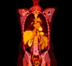
March 5, 2008 – Combining PET and CT for radiation therapy treatment planning in head and neck carcinoma patients provides local and regional disease control in comparison to CT alone, said a study in the March 1 issue of the International Journal for Radiation Oncology Biology Physics, the official journal of the American Society for Therapeutic Radiology and Oncology.
This study was conducted to calculate the clinical outcomes, including overall survival, disease-free survival and the incidence of recurrence of patients receiving PET/CT-guided radiation therapy and the correlation of the clinical outcomes to the maximum standard uptake value obtained on the PET scan.
According to the study, between Dec. 2002 and Aug. 2006, 42 patients with a median age of 55, who were diagnosed with head and neck squamous cell carcinoma, were given PET/CT imaging as part of their radiation therapy planning. The patients were monitored for at least six months following their treatment, with a mean follow-up time of 32 months.
Overall survival of the 42 study patients was 82.8 percent at two years and 74.1 percent at three years, superior to the survival rate that was found in a Radiation Therapy Oncology Group study in which patients received standard fractionation or accelerated fractionation with concomitant boost, said the study.
Results found that disease free survival for the 42 study patients was 71 percent and 66.9 percent at two and three years respectively. The cumulative incidence of recurrence was 18.7 percent. The study also found that standard uptake value quality predictor of local recurrence and that dose escalation based on standard uptake value is unlikely to be a fruitful treatment strategy, said the organization.
CT has been the go to decision for treatment planning for head and neck squamous cell carcinomas, but PET is proven to have an advantage over CT and other imaging modalities for primary tumor detection, including involved lymph nodes and distant metastatic disease not clearly otherwise identified. Using solely PET does have several disadvantages though, such as poor correlation to precise anatomic structures, but these negative impacts are reduced when PET and CT are combined by fusing the separate scans taken on a hybrid scanner.
For more information: www.astro.org

 April 23, 2026