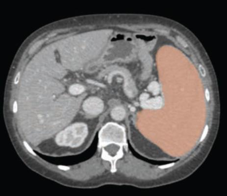
60-year-old woman with cirrhosis and portal hypertension, who underwent contrast-enhanced CT as pre-liver transplant evaluation. Patient weight was 74.4 kg. Automated deep-learning artificial intelligence tool was used to segment spleen and thereby compute splenic volume. Axial image shows spleen segmentation (orange overlay). Automated splenic volume was 1,097 ml, above weight-based splenic volume threshold for determining splenomegaly of 350 ml. True-craniocaudal splenic length was 15.5 cm, and maximum-3D splenic length 18.7 cm. These length measurements would indicate presence of splenomegaly at all thresholds used.
June 29, 2023 — According to an accepted manuscript published in ARRS’ own American Journal of Roentgenology (AJR), using an automated deep-learning AI tool, as well as weight-based volumetric thresholds, might afford large-scale evaluation for splenomegaly on CT examinations performed for any indication.
Noting that, historically, the standard linear splenic measurements used as a surrogate for splenic volume yielded suboptimal performance in detecting volume-based splenomegaly, “the weight-based volumetric thresholds indicated the presence of splenomegaly in most patients who underwent pre-liver transplant CT,” explained corresponding author Perry J. Pickhardt, MD, from the department of radiology at University of Wisconsin School of Medicine & Public Health.
Pickhardt and colleagues’ AJR accepted manuscript included a screening sample of 8,901 patients (4,235 men, 4,666 women; mean age, 56 years) who underwent CT colonoscopy (n = 7736) or renal-donor CT (n = 1165) from April 2004 to January 2017. A secondary cohort of 104 patients (62 men, 42 women; mean age, 56 years) with end-stage liver disease underwent pre-liver transplant CT from January 2011 to May 2013. Pickhardt et al.’s deep learning algorithm—previously developed, trained, and tested at the National Institutes of Health Clinical Center—was used for spleen segmentation, to help determine splenic volumes, with two radiologists independently reviewing a subset of said segmentations.
Ultimately, this automated deep-learning AI tool was utilized to calculate splenic volumes from CT examinations in 8,853 patients from the primary outpatient population. Additionally, splenic volume was most strongly associated with weight, among a range of patient factors.
“To our knowledge,” the AJR authors concluded, “this study represents the largest reported sample of patients to undergo volumetric segmentation of the spleen.”
For more information: www.arrs.org

 July 16, 2026


