
Aug. 8, 2025 — Memorial Hermann-Texas Medical Center announced it will begin using a new FDA-approved ultrasound device ...
Ultrasound Imaging
Ultrasound uses sound waves to create images. This section includes breast ultrasound, echocardiography (echo), transthoracic echo (TTE), transesophageal echo (TEE), echo contrast, transducers, ultrasound software and point-of-care ultrasound (POCUS).

July 24, 2025 — Fujifilm Sonosite, Inc., a leader in point-of-care ultrasound (POCUS) solutions, has announced a new ...
 July 24, 2025
 News | Ultrasound Imaging
July 14, 2025 — Patients with liver diseases will have expanded access to advanced ultrasound imaging and transplant ...
July 14, 2025 Sponsored Content
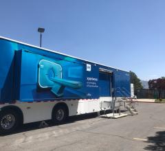
Case Study | Radiology Imaging
In June, the Philips Radiology Experience Tour hit the road to provide healthcare professionals with an opportunity to ...
September 19, 2023 
News | FDA
July 8, 2025 — Mendaera, Inc., a healthcare technology company focused on developing robotics that can be deployed ...
July 08, 2025 
News | Ultrasound Imaging
July 1, 2025 — UPDATE: The final paper is now available at: JMIR AI - ChatGPT-4–Driven Liver Ultrasound Radiomics ...
July 01, 2025 
News | Ultrasound Imaging
June 26, 2025 — Fujifilm VisualSonics Inc., a provider of ultra-high frequency ultrasound and photoacoustic imaging ...
June 27, 2025 Sponsored Content

News | Radiology Imaging
January 23, 2023 — Canon Medical Systems has released a new eBook featuring its new medical imaging roadshow. This new ...
January 23, 2023 
News | Ultrasound Imaging
June 16, 23025 —GE HealthCare has launched the bkActiv S series, representing the next era of intraoperative ultrasound ...
June 25, 2025 
June 17, 2025 — Royal Philips has announced the global launch of the Flash Ultrasound System 5100 POC — a new point-of ...
June 19, 2025 
News | Lung Imaging
June 18, 2025 — Exo recently announced that now included on its Exo Iris is the first ever FDA 510(k) cleared AI for ...
June 18, 2025 Sponsored Content

Case Study | Ultrasound Imaging
The most common cause of chronic liver disease? Nonalcoholic fatty liver disease (NAFLD). With 25% of the world’s ...
January 19, 2022 
News | Ultrasound Imaging
June 4, 2025 — RadNet, Inc., a provider of high-quality, cost-effective diagnostic imaging services and digital health ...
June 09, 2025 News | Ultrasound Imaging
May 14, 2025 — A comprehensive new study based on nationwide claims data from more than 11 million patients shows that ...
May 14, 2025 
News | Artificial Intelligence
April 16, 2025 — An artificial intelligence (AI) program trained to review images from a common medical test can detect ...
April 16, 2025 Sponsored Content

Case Study | Ultrasound Imaging
There is a high incidence of ultrasound work-related musculoskeletal disorders (WRMSD) among sonographers. Aside from ...
November 12, 2020 
News | Ultrasound Women's Health
April 11, 2025 — Contrast-enhanced ultrasound (CEUS) is a safe and accurate diagnostic imaging option for pregnant women ...
April 11, 2025 
News | Ultrasound Imaging
March 31, 2025 — MedStar Georgetown University Hospital is the first hospital in the Washington, D.C. region to offer ...
April 02, 2025 
News | Focused Ultrasound Therapy
March 31, 2025 — Neuropathic pain affects up to 10 percent of the global population and can be challenging to manage ...
April 02, 2025 
News | Breast Imaging
March 20, 2025 — GE HealthCare has launched Invenia Automated Breast Ultrasound (ABUS) Premium, the latest 3D ultrasound ...
March 21, 2025 
News | Ultrasound Imaging
March 13, 2025 — Vave Health has introduced its Universal Wireless Probe. The probe is designed to enhance efficiency ...
March 20, 2025 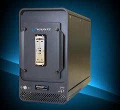
News | Ultrasound Imaging
March 20 — Verasonics, Inc. recently announced the release of new features for the Vantage NXT Research Ultrasound ...
March 20, 2025 
News | X-Ray
March 18, 2025 — GE HealthCare recently announced a collaboration with NVIDIA expanding the existing relationship ...
March 19, 2025 
News | Breast Imaging
Feb. 26, 2025 — iCAD, Inc. a provider of clinically proven AI-powered cancer detection solutions, and Koios Medical, a ...
March 03, 2025 © Copyright Wainscot Media. All Rights Reserved.
Subscribe Now