
Sept. 13, 2024 — Bayer Calantic Digital Solutions has announced the availability of a new eBook that addresses how ...
Artificial Intelligence
This channel includes news and technology innovations for artificial intelligence (AI) software, also referred to as deep learning, cognitive computing and machine learning. AI technology is being integrated in radiology for imaging appropriate use criteria (AUC), clinical decision support, predictive analytics and to assist radiologists with improved workflow.
News | Breast Imaging
Aug. 28, 2024 — Rezolut, LLC recently debuted its latest offering for patients during their annual mammogram ...
 August 29, 2024
News | Digital Pathology
AI Tool Simultaneously Screens 505 Genes for Comprehensive Cancer Diagnosis, Personalized Treatments
Paige has launched OmniScreen, an AI-driven biomarker module capable of evaluating over 505 genes and detecting 1,228 ...
August 27, 2024 Sponsored Content
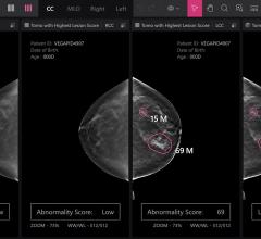
Feature | Breast Imaging
Despite decades of progress in breast imaging, one challenge continues to test even the most skilled radiologists ...
October 24, 2025 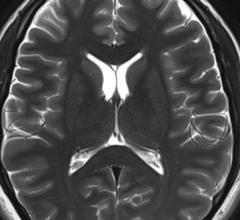
News | Artificial Intelligence
Aug. 26, 2024 — Data scientists and clinical researchers will use brain scans from the entire Scottish population to ...
August 26, 2024 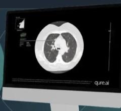
News | Artificial Intelligence
Aug. 23, 2024 — Qure.ai recently announced its AI-powered chest CT solution, qCT LN Quant, has received 510(k) FDA ...
August 23, 2024 
News | RSNA
July 31, 2024 — The National Imaging Informatics Course (NIIC), a pioneering program in the radiology field, will return ...
July 31, 2024 Sponsored Content
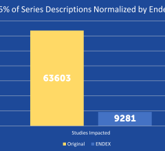
News | Artificial Intelligence
SPONSORED CONTENT — EnsightTM 2.0 is the newest version of Enlitic’s data standardization software framework. Ensight is ...
June 21, 2024 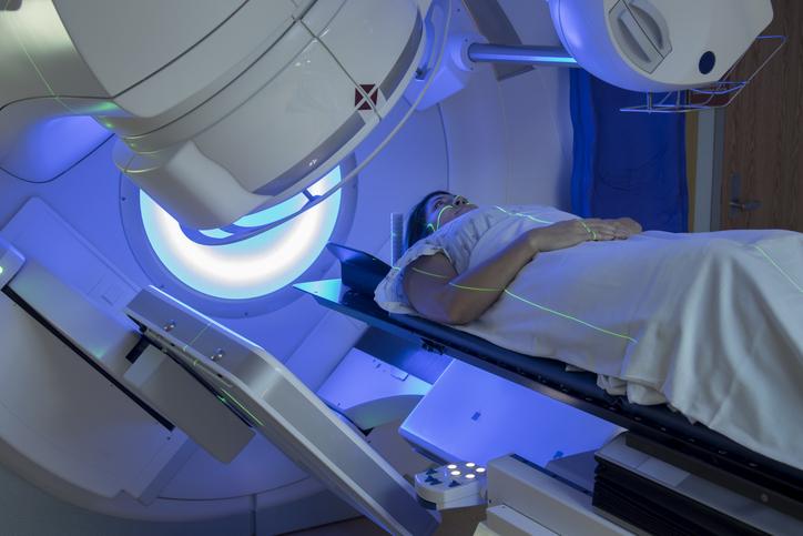
Feature | Radiation Oncology | By Christine Book
News emerging from several leading organizations and vendors in the radiation therapy arena came in at a fast pace in ...
July 30, 2024 
News | Breast Imaging
July 29, 2024 — Lunit, a leading provider of AI-powered solutions for cancer diagnostics and therapeutics, announced the ...
July 29, 2024 
News | Breast Imaging
July 29, 2024 — iCAD, Inc., a global leader in clinically proven AI-powered cancer detection solutions, announced a ...
July 29, 2024 Sponsored Content

Feature | Artificial Intelligence
Did you know that approximately one-third of all the data in world is created by the healthcare industry and that ...
June 03, 2024 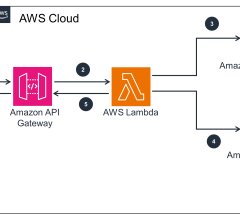
News | Artificial Intelligence
July 26, 2024 — GE HealthCare and Amazon Web Services, Inc. (AWS), an Amazon.com, Inc. company, announced a strategic ...
July 26, 2024 
Videos | Information Technology
Industry trade shows and conferences seem to be making their comeback in 2024. And the Healthcare Information and ...
July 25, 2024 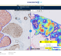
News | Digital Pathology
July 24, 2024 — Proscia, a developer of artificial intelligence (AI)-enabled digital pathology solutions for precision ...
July 24, 2024 Sponsored Content

Case Study | Enterprise Imaging
Having the most efficient clinical workflows with enhanced diagnostic capabilities is a major goal for clinicians and ...
May 16, 2024 
Videos | Breast Imaging
Don't miss ITN's latest "One on One" video interview with AAWR Past President and American College of Radiology (ACR) ...
July 24, 2024 
News | RSNA
July 23, 2024 — Professional registration is open for RSNA 2024, the world’s largest radiology forum. This year’s theme ...
July 23, 2024 
News | Artificial Intelligence
July 23, 2024 — Researchers at the National Institutes of Health (NIH) found that an artificial intelligence (AI) model ...
July 23, 2024 
News | Artificial Intelligence
July 22, 2024 — Healthcare artificial intelligence (AI) systems provider, Qure.ai, has announced its receipt of a Class ...
July 22, 2024 
News | Artificial Intelligence
July 17, 2024 — Hyperfine, a groundbreaking medical device company that has redefined brain imaging with the world’s ...
July 17, 2024 
Feature | Imaging Technology News - ITN
Be sure to check out the latest digital edition of Imaging Technology News (ITN), featuring the Mobile C-arm Systems ...
July 11, 2024 
News | Prostate Cancer
July 11, 2024 — GE HealthCare’s MIM Software, a global provider of medical imaging analysis and artificial intelligence ...
July 11, 2024 
News | Artificial Intelligence
July 11, 2024 — Artificial Intelligence (AI) tools can play a key role in medical imaging if radiologists trust in their ...
July 11, 2024 © Copyright Wainscot Media. All Rights Reserved.
Subscribe Now