Would accessibility from anywhere at the expense of basic visualization tools offered by a thin-client configuration satisfy your radiology needs? Or would you rather have the full functionality a slim client supports at the cost of more bandwidth? You make the call. Or can you have the best of both worlds?
There is a new twist on the slim- vs. thin-client debate. You may be able to utilize a slim client without buying more bandwidth. Or you may consider recently released thin-client upgrades with more robust tool sets.
How thick is slim?
The minimal bandwidth requirement for a slim-client is 100 MB, but according to Terry Chang, director of product management at Ziosoft, this is not such a tall order. “Most hospitals and clinics meet this requirement,” noted Chang. “Utilizing higher network bandwidth, greater than 100mb/s, across their facilities, makes the slim client a good choice for enterprise accessibility of advanced visualization.”
But what about the off-site radiologist or referring physician? Is it worth it to beef up the bandwidth to go from thin to slim?
Before investing in more bandwidth, there are a few things to consider. In both thin- and slim-client configurations, there is software on both the client workstation and server as opposed to a stand-alone workstation that has all the software on the workstation without a server. Most of the “heavy lifting” image processing is performed on the server. This centralized processing scheme is designed to improve efficiency and promote increased accessibility for multiple users. For both configurations, it’s important to note if any proprietary hardware is required, this will affect cost and scalability.
What distinguishes the two solutions is the available toolset and the network. “The difference in thin versus slim comes down to level of required functionality and available network bandwidth,” explained Chang. “Thin-clients require less bandwidth, while slim-clients require more bandwidth. As a result, in a thin client, performance is limited and more sophisticated functionality, such as real-time automatic segmentation and fly-through, are usually not possible. In this case, a separate stand-alone workstation is required for cases that require full functionality; however there is usually a difference in the user interface between a stand-alone workstation and thin-client application. This can be a source of frustration for users if they have to utilize both.”
Since thin-client performance is limited, the required software on the client can be ‘thinner’ than a slim client. “The ‘thinnest’ thin client would be a Web browser-based application,” said Chang, “that requires very little if any software on the client workstation, but functionality would probably be further limited.”
The slim client/server architecture enables radiologists to perform full-functional advanced visualization in any location throughout the enterprise. Radiologists do not have to use two separate systems for basic and sophisticated volume rendering. On the Ziostation application, a product developed by Ziosoft Inc., ‘intelligent’ tools such as action macros, automated curved planar reconstruction and automatic segmentation — in other words, bone removal and vessel extraction - are available. Since the ZIOSTATION utilizes all standard commercial hardware, PACS integration capability is also optimized, said Chang.
Centralized and consistent studies promote good collaboration amongst other clinicians too. The type of users who would benefit from a thin client are ones that do not have access to higher speed networks and accept not having all the sophisticated functionality of a stand alone or slim client. These users, for example, referring physicians and users who need access in remote locations such as from home or while traveling, require only basic functionality and the ability to simply view advanced visualization images.
Is thin catching up to slim?
With the slim-client offering a better toolset, it seems obvious to set up a slim-client solution if you can afford the bandwidth. But not so fast. Thin might be catching up to slim, in a move that Visage Imaging calls ‘thinnovation.’
While most thin-client concepts are simply an extension of a traditional workstation to allow for remote usage, other players, like Visage, Terarecon and Vital Images claim they go beyond these basic offerings.
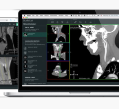
Visage Imaging’s Visage Thin Client solutions include advanced tools for 2D, 3D and 4D image review and interpretation, post-processing, data management and image distribution. The company says that one of the key concepts behind its thin client is true integration within the RIS/PACS workflow or enterprise-wide diagnostic imaging workflow. Visage also claims to provide the full functionality that is needed in the imagery-based workflow provided within the thin-client platform. Its most recent upgrades reportedly offer application-specific display and post-processing protocols, saving and sharing of annotations as well as post-processing results, volume analysis of lesions and structures in 3D, improved automatic bone removal, sharing of roaming sessions, easy switching of layouts and viewers across radiology, cardiology, neurology, oncology, surgery and other subspecialties.
“The key differentiator of a thin-client PACS – a PACS with tightly integrated 3D thin-client technology – is a true central 3D processing paradigm, along with efficient streaming technology to enable thin clients to act as fully capable front-ends to all viewing and processing functions,” said Gabi Strasser, marketing and communications manager, Visage Imaging, Visage Imaging GmbH. “All DICOM data remains on the server, meaning that no data transfer is necessary prior to launching the 3D viewer and the images are instantly available. All operations are performed directly on the server from anywhere in the hospital at the speed that only a full-blown server machine can provide.”
Visage Imaging will soon roll out a new version of Visage CS Cardiac Analysis, which will feature a reportedly more robust set of tools for calcium scoring, improved reporting and efficient manual editing of the left ventricle geometry. Visage CS Cardiac Analysis will allow viewing and post-processing of even the largest multiphase cardiac CT studies.
Centralize the “heavy lifting”
The key to augmenting the power of either a slim or thin client is doing the “heavy-lifting” of performing the volume rendering work on centralized servers that are far more powerful than each individual workstation.
If the rendering is performed on a large server and the resultant images are streamed back to the user in an interactive fashion, it creates a real-time workstation experience. This allows for the workstation to be anywhere, and for the user to work on a low-end PC system or a fully loaded radiology review station.
TeraRecon’s AquariusAPS is designed to provide greater automation by carrying out many zero-click tasks before the radiologist opens the study. If a technologist is preparing the study first, every image they store is a saved state that can be further manipulated and validated without limitation. Upon reading the first study of each type, Aquarius iNtuition learns the reading flow and creates a volumetric 3D hanging protocol called a “workflow scene” and future studies received by the system can be hung in this same way automatically according to the preferences of every user.
Souped up toolset
So how far can thin client soup up its advanced visualization toolset and match that of a slim client?
Take the Aquarius iNtuition platform’s toolset, which is sizeable for a thin-client offering optional clinical tools such as soft plaque analysis, CT/PET fusion, volumetric subtraction (dual energy), enhanced DICOM support (320/256 slice CT scanners support), EP planning, conferencing and collaboration.
Vital Images’ latest next-generation release, ViTALConnect 4.1 Web-client, includes enhancements with new cardiovascular tools, such as automatic segmentation of the coronary tree, centerline editing and automatic stenosis calculation. Its Vitrea 4.0 workstation-client upgrade also provides those features ViTALConnect 4.1 does, plus probing of the coronary tree, vessel management and labeling and reporting with automatic population of findings.
Vitrea 4.0 adds a few neurovascular improvements that include motion correction for true 4D perfusion for stroke patients, plus for colon, fly-through, polyp probe and automatic registration of prone and supine, and CAD for colon.
“What I demonstrated live at the recent Stanford course — loading and analyzing a 5,020 slice study over the Web-client — I could have done from my house using my laptop,” said Tony DeFrance, M.D., medical director of the CVCTA Education Center in San Francisco. “I routinely use the workstation client, but it’s nice to know that I can use the same powerful tools remotely using the Web client.”
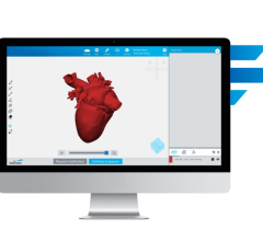
The new EP planning application, ViTAL EP, contains a 3D advanced visualization and modeling tool for the electrophysiology (EP) lab. ViTAL EP is said to automatically segment out the pulmonary veins and creates a three dimensional anatomic model of the heart for super-imposed EP mapping. In addition to its powerful clinical features, ViTAL EP automatically exports a 3D model for display on the St. Jude Medical EnSite System, which is used to facilitate therapy for the treatment of arrhythmias.
While the appropriateness of a slim- or thin-client application is very individual, your decision will depend on a few key factors: what level of functionality you need, how much bandwidth you have, and if your servers can be clustered to leverage more power. And, of course, always remember cost, which more than anything may depend on your negotiating power.


 April 18, 2025


