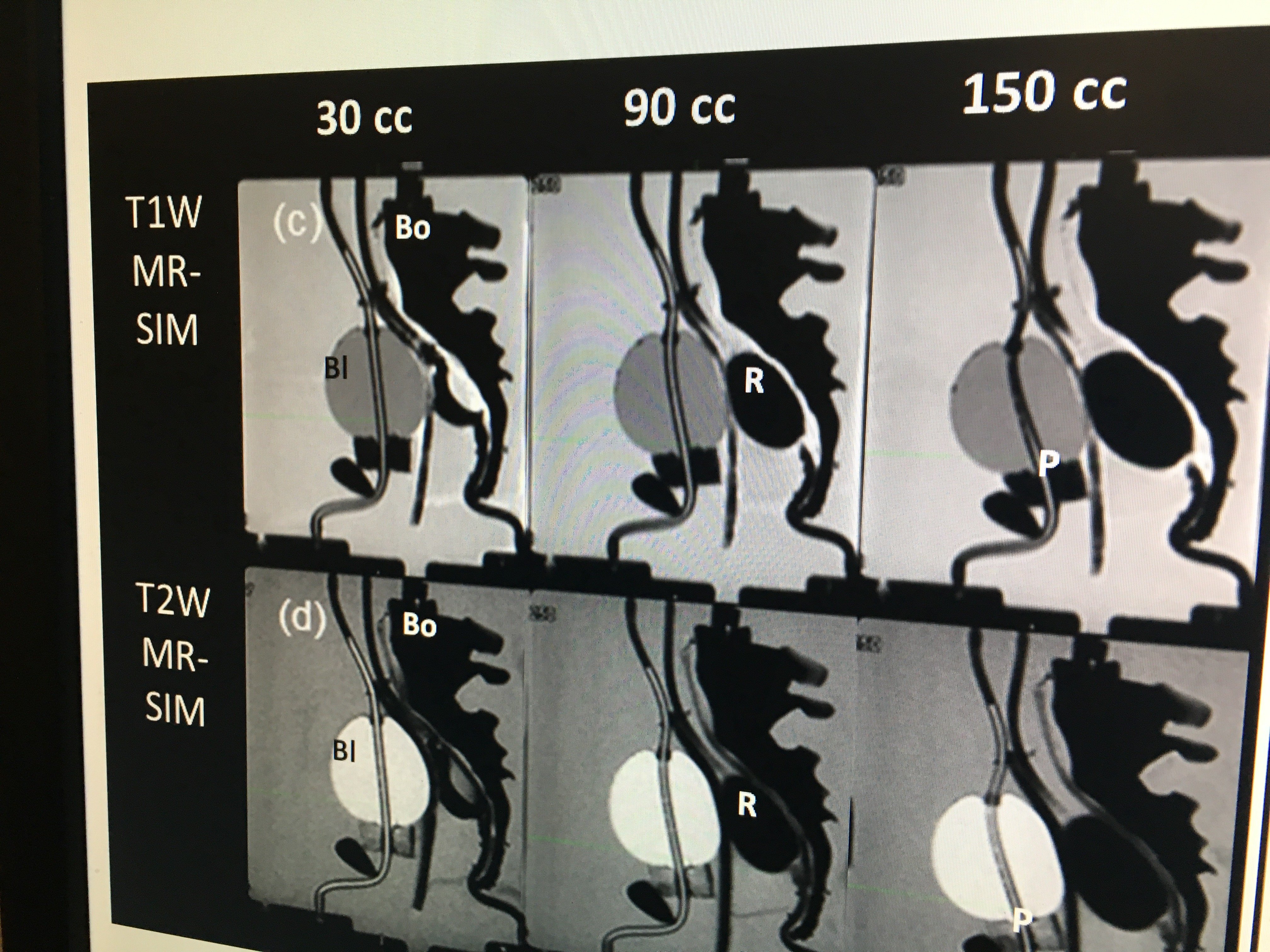
A set of synthetic CT images created from T1 and T2 weighted MR imaging of a prostate/rectum phantom at Henry Ford Hospital. The hospital is one of the research centers developing synthetic CT imaging for treatment planning to avoid the need for CT scans of a patient just for treatment planning purposes when they already have a more detailed soft tissue MRI exam of the anatomy.
Computed tomography (CT) has traditionally played an important role in the development of radiation therapy treatment plans for cancer patients. The Hounsfield unit measurements acquired from a CT scan are critical for therapeutic dose calculations to ensure that the most dose is delivered to the target region, while sparing healthy tissue as much as possible. Aside from dose calculation, however, CT has inherent limitations that require the use of other modalities like magnetic resonance imaging (MRI) to generate a complete, optimal treatment plan.
In these scenarios, the two image sets must be coregistered in order to display the most complete picture with all of the information needed for planning. Coregistration comes with its own set of uncertainties, however, over the images not being aligned correctly. Moreover, this methodology still utilizes CT, exposing the patient to even more radiation dose when they are already receiving a large therapeutic dose.
It is for these reasons that clinicians and vendors throughout the industry have been looking for new imaging methods that might resolve all of these issues. One avenue that has shown promise in recent years is the creation of synthetic CT images from an MR-only imaging sequence. “It’s more efficient, [with] fewer things to contour and coregister, so we expect coregistration uncertainty to be reduced by having only one imaging modality, which is the one that is most desirable for a lot of disease sites,” said Carri Glide-Hurst, Ph.D., director of translational research, radiation oncology at Henry Ford Health System in Detroit.
New Research Arenas in Synthetic CT
Glide-Hurst and her colleagues at Henry Ford have spent the last several years exploring new applications for synthetic CT in treatment planning, as well as ways to enhance the utility of the technology. One area they have focused on in particular is deriving the CT Hounsfield units needed for therapeutic dose calculation from MRI-based synthetic CT.
Glide-Hurst credited junior physicist Joshua Kim, Ph.D., for a prostate cancer-specific solution to enhance synthetic CT dose calculation. “Essentially what it is, our prostate solution relies on a T1 and T2 input dataset, and then it’s a voxel-based weighted summation and it’s got patient-specific output. It gives a very good agreement to the CT both in the bones and the tissue and all of the different soft tissue,” Glide-Hurst said.
The Henry Ford group has also taken interest in using synthetic CT for brain cancer treatment planning, particularly for image-guided radiation therapy (IGRT), where the application is not as well understood. “The brain is a little trickier because we have to worry about some small bones and small structures, and a much more complex anatomy there. Accuracy is very, very important in the brain,” said Glide-Hurst.
A 2018 study by the group compared geometric equivalence of digitally reconstructed radiographs (DRRs) from CTs and synthetic CTs to quantify performance for partial brain image-guided radiation therapy (IGRT). For the study, 10 brain cancer patients underwent both MRI and CT simulation. Synthetic CTs were generated by combining ultra-short echo time, T1, T2 and fluid attenuation inversion recovery datasets using voxel-based weighted summation. The two types of DRRs were compared using patient-specific thresholding and assessed using:
• Overlap index;
• Dice similarity coefficient; and
• Jaccard index.
Planar IGRT images for 22 fractions were evaluated for differences between the two DRR types in six quadrants. Ultimately, statistically significant differences between CT and synthetic CT registrations were observed in 9 of 18 matched quadrants/axes. The researchers concluded that while the DRR synthetic CT geometry was robust, the results were not clinically significant.1
Commercial Synthetic CT Products
As the technology continues to prove valuable, vendors from both the imaging and treatment planning sides have begun releasing their own synthetic CT products.
Siemens Healthineers introduced synthetic CT capabilities on the Magnetom RT Pro edition of the Magnetom Sola MRI scanner, debuting at the 2018 American Society of Radiation Oncology (ASTRO) meeting. The 1.5T scanner has multiple new features tailored specifically for treatment planning, highlighted by RT Image Suite, a dedicated post-planning software. Synthetic CT capabilities of the RT Image Suite address five major areas:
• Imaging for planning and simulation: The
scanner’s RT Dot Engine provides workflow
guidance during image acquisition, with
dedicated RT protocols for brain, head and
neck, and pelvis exams.
• Post-processing and assessment: After
acquisition, the synthetic CT image is
automatically created and displayed on the
scanner console.
• Contouring: The system displays the synthetic
CT alongside other MR images for patient
marking, assessment and parallel contouring.
Once this step is complete, the synthetic image
can be exported to the treatment planning
system for dose calculation.
• Dosimetric planning: Users can correlate the
synthetic CT dose calculations with
conventional CT to check agreement.
• Treatment monitoring and adaptation: Siemens
included a variety of applications that can aid
in assessing treatment response alongside
synthetic CT images, including MR OncoCare
for functional characteristics of a lesion.
On the treatment planning side, Sweden-based Spectronic Medical received European CE mark in 2016 for its MRIPlanner software. The software facilitates the creation and transfer of synthetic CT images between the MRI scanner and the treatment planning system (TPS). A LinkClient DICOM node installed at the hospital receives the original MR images from the scanner and forwards them to the MRIPlanner software. Upon completion, the node returns the synthetic image with the proper delineations to the TPS.
A 2017 study published in the International Journal of Radiation Oncology Biology Physics demonstrated that the MR-only workflow of MRIPlanner was dosimetrically accurate and robust for a variety of equipment vendors, magnetic field strengths and treatment techniques. Mean dose differences between the CT and synthetic CT images in the MR-OPERA study were below 0.25 percent for all evaluated organ and target structures; gamma evaluation showed a mean pass rate of 99.12 percent in complete body volume and 99.97 percent in the planning target volume. Results were consistent across the four centers participating in the study.2
The first 30 clinical patients were treated with MRI Planner in January 2018. Spectronic Medical said that research is ongoing to explore applications for MRIPlanner in brain and head and neck cancer cases. The software has yet to receive U.S. Food and Drug Administration (FDA) approval.
References
1. Morris E.D., Price R.G., Kim J., et al. Using synthetic CT for partial
brain radiation therapy: Impact on image guidance. Practical
Radiation Oncology, September-October 2018. Published first
online April 6, 2018. https://doi.org/10.1016/j.prro.2018.04.001
2. Persson E., Gustafsson C., Nordström F., et al. MR-OPERA: A
Multicenter/Multivendor Validation of Magnetic Resonance
Imaging–Only Prostate Treatment Planning Using Synthetic
Computed Tomography Images. Red Journal, June 15, 2017
https://doi.org/10.1016/j.ijrobp.2017.06.006
Related content:
Find more radiology and radiation oncology content from Henry Ford Hospital

 June 04, 2026


")

