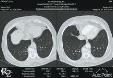
R2 Technology integrated its ImageChecker CAD CT Lung solution into Vital Image's Vitrea workstation to optimize workflow demands of lung examiniations
The good news is that hybrid imaging, one of the fastest-growing markets in medical imaging, is an effective tool for noninvasive disease detection, monitoring and therapy. The trouble is that many physicians cannot optimize the use of all the high-volume data generated because their workstations lack advanced visualization applications for rendering 3-D and 4-D images.
As more physicians today regularly switch viewing between 2-D, 3-D and 4-D images, workflow can be significantly hindered when switching between applications located at separate clinical workstations or on a semi-integrated system. Adequate workstations today must not only manage large volume data sets but also come equipped with fully integrated 3-D and 4-D post-processing applications accessible across the enterprise and remotely.
But a generic 3-D solution is not enough – the need for advanced visual applications goes beyond the radiology department, as oncologists, neurologists and cardiologists are also routinely consulting images produced from PET, SPECT, CT and MR. The good news is that PACS vendors are catching on and designing specialty applications for specific focuses, such as colon, cardiology, lung, breast and bone that incorporate 3-D and 4-D into clinical workflow – replacing generic 3-D applications.
Hybrid Imaging Grows 3-D, 4-D
Hybrid imaging is among the fastest growing diagnostic imaging sectors and is also driving the demand for multiple applications on a single workstation.
Playing a unique role in disease detection, fused images provide detailed anatomy and functional processes at the molecular level, allowing physicians to noninvasively pinpoint the exact location, size, nature and extent of disease anywhere in the body. In addition to disease detection, hybrid imaging is increasingly used to track therapy, detecting whether a specific chemotherapy is effective for an oncology patient or how fast the chemotherapy is working. As a result, physicians are routinely viewing hybrid images rendered on 2-D, 3-D and 4-D applications.
“As the growing number of high-volume CT procedures mandates the use of advanced visualization tools, radiologists require a workstation set up for simultaneous viewing of 2-D, 3-D and 4-D images,” said Eliot L. Siegel, professor of Diagnostic Radiology, director, Baltimore Veterans Affairs Medical Center Radiology, associate vice chairman for Informatics.
Fully Integrated for Full Efficiency
Rising volumes of multimodality images bring with them “CT data overload,” as Dr. Siegel calls it. To handle all of this data without hampering productivity, workstations need advanced visualization tools that are fully integrated into the PACS.
“Advanced visualization software including 3-D and 4-D capabilities are, in my opinion, essential for routine interpretation of all imaging studies and should be part of the image interpretation process,” said Siegel. “When we refer to a PACS workstation, we’re typically referring to the workstation that is used routinely for retrieval of a reading worklist and has prior studies and reports and the indication for a current study. In order to have an efficient workflow, it is much better to have advanced visualization capability integrated with the PACS than as a stand-alone or poorly integrated application.”
Consider something as subtle as the location of image display; the position of displays can significantly help or hamper workflow. By integrating the radiologist’s toolset — 2-D, 3-D and 4-D applications with PACS — workflow is made faster and easier. Many workstations have semi-integrated visualization tools to allow radiologists to open a study in a 3-D application without using a worklist, but unless these applications are fully-integrated into a PACS, any changes that are made in the 3-D viewer are not duplicated in the 2-D system in the PACS. Like a PDF, once 3-D models are created, the 3-D image is for static viewing only. This results in decreased efficiency.
To better streamline workflow, solutions like Aquarius Workstation by TeraRecon offer a fully-integrated suite of clinical application modules and enable real-time diagnostic review of 2-D, 3-D and 4-D images for large-slice CT and MR.
With Merge eMed’s 3-D/4-D Review application comprehensive visualization capabilities are available for rapid and efficient everyday clinical workflow along with a real-time editing tool in combination with standard visualization tools such as MIP, MinIP and 3-D VRT. Users can reportedly visualize the removal of overlaying structures on-the-fly.
Another opportunity to increase efficiency is through modality-specific tools where users set the protocols and do not need to search the desktop. This can be achieved through a digital cockpit or dashboard that integrates images and data from multiple stand-alone applications into a common display. Cedara offers C4 (Cedara Clinical Control Center) to help manage multiple plug-ins, including deployment, auto-launching, as well as management of input and outputs.
Specialized 3-D, 4-D Applications
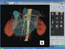
Viewing high-volume 3-D and 4-D data sets is becoming prevalent across specialties, and specific applications for colon, cardiology, lung, breast and bone are now incorporating 3-D and 4-D into clinical workflow.
TeraRecon recently upgraded Aquarius Workstation version 3.5, which offers protocol-based workflow tools that automatically present the 3-D volume data set based on the study type selected by the operator. For cardiovascular CT for example, the software offers new soft plaque analysis, automatic vessel segmentation and extraction, measurement, interpretation and reporting tools. For CT colonography, the entire colon is automatically segmented upon loading with flight paths automatically generated, enabling the user to switch from 2-D synchronized views to 3-D fly-through mode.
Engineered for integration with advanced visualization and clinical imaging applications, FUSION RIS/PACS by Merge includes 3-D/4-D post-processing for virtual colonoscopy, CT angiography, breast imaging, lung nodule detection, calcium scoring, image stitching and more.
Server-Based Viewing – Anytime, Anywhere
The need to view high-volume images from any location in a hospital is becoming more ubiquitous as physicians from various disciplines are using multimodality imaging and are frequently consulting across specialties. This requires the use of 3-D and 4-D applications anywhere at anytime. Because server-based applications enable enterprise-wide and remote access to high-volume images, without the need to purchase software licenses or complex hardware upgrades at each location, they may set the new standard.
“Large volume data sets required for 3-D and 4-D image review will force a change from client-based to server-based image rendering in order to minimize the time required for image review, to maximize the power available at the client workstation, to keep costs reasonable and to eliminate the need to upgrade both hardware and software on PC’s throughout the imaging department and the enterprise,” noted Siegel.
One new server-based application is AquariusNET Server version 1.7 by TeraRecon, which routes CT slices for immediate interactive 3-D review by radiologists and clinicians from regular client PCs anywhere throughout the enterprise. For advanced 3-D analysis, CT and MR scans can be sent to the Aquarius Workstation where the technologists or physician can perform initial processing, removing bone and nonrelevant structures or segmenting vessels from surrounding tissue. The images are then stored on the server so that physicians may open the exam and pick up where the previous clinician left off. The original axial images, interactive 3-D and MIP/MPR reformats are backed up on the server. For offsite access, TeraRecon offers AquariusNET thin-client application, which is reportedly compatible with most PACS diagnostic review stations, HIS, RIS and other informatics systems.
“Seamless connectivity across our network allows us to fully leverage our physicians’ subspecialty expertise and get the right images to the right physician regardless of where they were generated and regardless of where across our network the physician may be located. We struggled to find the right IT consultants that could look at so many different sites using so many different PACS/RIS systems and to link them into an efficient operating network,” said John Anastos, M.D., department of radiology chairman at Advocate Lutheran Hospital and president of ARC, SC.
ARC chose software developer Compressus to link its medical imaging and data management systems at 11 ARC sites. To integrate disparate information systems and enable users to select best-of-breed PACS, HIS/RIS, EMS and CIS tools, Compressus implemented MEDxConnect System, a server-based open-architected software that created a virtual archive and database for high-speed enterprise-wide data transfer from any acquisition device to any workstation.
Innovation Creates New 3-D Applications
Necessity is the mother of all invention, including new clinical applications for 3-D imaging. One recent technological innovation that will create new uses for 3-D is a new method to acquire volume dual-energy images, which was performed on a GE Healthcare CT system by Sachio Kuribayashi, M.D., chairman and professor of Radiology, Keio University in Japan. “This approach, utilizing a unique acquisition protocol useable to the full 50cm field of view, may help clinicians expand clinical capabilities with new applications that could include material decomposition – separation of iodine and calcium and simplifying bone vessel segmentation,” said Gene Saragnese, global vice president of Molecular Imaging and CT, GE Healthcare. While GE is currently investigating what clinical applications may benefit from this technology, such as separating calcium and iodine, improving segmentation of vessels and bone particularly in the base of skull, or identifying iron in the liver, GE is considering “how to best display these images and 3-D rendering is one area,” according to Jamie Valliant, marketing manager, CT Advanced Technology Programs, GE Healthcare.
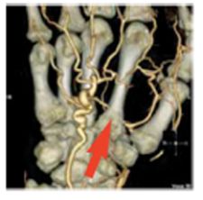
Significant advances have also been achieved in segmentation technologies, which allow clinicians to remove extraneous anatomy during review, but one of the more difficult segmentation challenges is separating small arteries from bone – for example, the deep palmar arch – and isolating this artery using traditional 3-D workstation software tools.
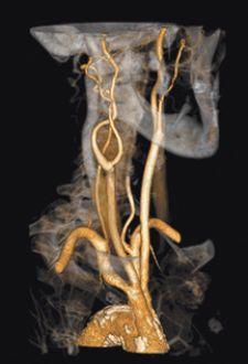
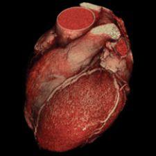
Barco just received FDA 510(k) clearance for CardiaMetrix, its clinical application suite for structural and functional analysis of contrast-enhanced cardiac studies. Integrated into PACS with Voxar 3D and VesselMetrix, a clinical application for quantitative vessel analysis of CTA and MRA, CardiaMetrix’s segmentation and bone removal software is designed to allow users to extract small vessels quickly and provides a 4-D cardiac analysis tool to produce 4-D movies of the beating heart with cardiac contours. The 4-D cine rapidly generates short axis, two-, three- and four-chamber long axis views, semi-automatic alignment with cardiac long axis position and automatic transfer of long axis position in end-diastolic and systolic phases to cardiac (LV) analysis.
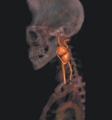
First-Time 4-D CT Angiography
A new concept called 4-D CT angiography (4-D CTA) may open up new clinical applications for angiography such as ‘dynamic blood flow’ and new uses for 4-D imaging. Professor Murakami, chairman, department of Diagnostic and Interventional Radiology at Kinki University Hospital, Japan, performed for the first time a dynamic CT angiography (CTA) and full organ perfusion study in one scan, reportedly without compromising physiological temporal resolution.
4-D CTA, realized on a GE Healthcare CT system, may allow clinicians to view in detail the relation between tumors and feeding arteries and determine the adequate treatment choice, ablation or medical therapy. The imaging technique may also mark a step toward breaking the barrier surrounding dynamic CTA and CT perfusion studies to create new clinical use in whole organ studies.
The challenge that physicians face today is to manage and optimize the use of high-volume data sets to create images that assist in making clinical diagnosis, tracking therapy and examining molecular activity to hopefully prevent disease before symptoms appear. To achieve this, clinical workstations must keep up with the evolution of technology, providing doctors with the imaging tools they need to render a higher level of care.


 April 18, 2025