Siemens' AXIOM Artis dTA C-arm scanning system
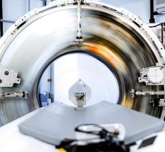
Anyone who remembers – or for that matter, still uses – single-slice CT scanners can appreciate the dramatic advancements made by leading CT system manufacturers in recent years. The latest CT, Multidetector CT (MDCT), and MRI systems are faster and feature higher resolution than ever before.
CT scanners alone are developing at breakneck speed. Siemens AG Medical Solutions now counts more than 350 installations for its 64-slice scanner, the Siemens SOMATOM Sensation 64. According to Stephen Mohan of consulting firm Frost & Sullivan’s North American Healthcare Practice, “healthcare professionals now consider [64-slice CT] an industry standard.” Challenging the medical imaging leaders such as GE Healthcare, Siemens and Phillips Medical Imaging, which have introduced their 64-slice CT scanners, earlier this year Toshiba Medical Systems presented a prototype of a 256-slice CT scanner at the American College of Cardiology meeting in Atlanta. A Toshiba representative estimated that the product would reach the market in just two years.
However helpful these new tools are in speeding diagnoses and improving accuracy, they are putting enormous pressure on the 3-D workstations designed to interpret imaging studies. The truth is, the type of volumetric information these systems generate is growing so rapidly that the visualization systems used in a variety of fields – medical research, manufacturing, even oil and gas exploration – are struggling to keep up. If the 3-D volume is too large for the workstation to handle, users have to view information in piecemeal, with system components swapping out one portion of an overall model with another.
Today’s workstations use optimized graphics technology, and this has helped. Unfortunately, the technology used in the systems often sacrifices quality in the rendering of the 3-D volume, and, in the medical field, any compromises in data accuracy and detail can be costly indeed.
A potential problem with these workstations is that they rely on something called a graphics processing unit (GPU). GPUs have been the engines that drive graphics in PCs and workstations for years, but the pace of GPU innovation has not kept up with the pace of scanner device innovation. Very soon, they will need help carrying the load.
No Room for Error
Fortunately, a rendering technology first developed 40 years ago is already being used by medical researchers and others to see volumes in exceptional detail. The technology is called real-time ray tracing. Ray tracing is a rendering technique that uses the behavior of light to generate extremely realistic, detailed images. Ray tracing software works by simulating a ray projected through a 3-D scene. It then calculates how the ray and the light source intersect with the objects in the scene. This results in renderings that present real-world 3-D volume data with photo-realistic accuracy – a breakthrough for imaging professionals whose work leaves little room for error.
It is fair to say ray tracing has its roots in the X-ray suite. It was invented to simulate the effects of nuclear penetration, but until the past few years, its use was limited. Ray tracing performs better when it runs on more processors (the “brains” of the computer, typically manufactured by Intel or AMD) and when it has access to more computer memory. Because GPUs are expensive and don’t include much memory, ray tracing never caught on with users of traditional PCs, laptops and workstations.
In recent years, however, a new generation of computers has arrived. These systems use powerful processors and make large amounts of computer memory available to the user. With computers like these, real-time ray tracing has been embraced in a broad range of environments, including medical research.
Real-time ray tracing is, at least in the medical field, still in its earliest stages, and it is likely to be three to five years before commercial visualization solutions incorporating the technology reach the market, and even those will likely be used by “early adopters.” But the work of some innovators offers a glimpse of its enormous promise.
Ray Tracing and Research
For these researchers, real-time ray tracing makes sense because their scanning and measurement devices are so advanced, generating up to 100 times the detail of traditional CT scanners. At the Lawrence Berkeley Laboratory (LBL), a “micron CT” scanner known as a Synchrotron records data at a resolution of 10 microns (compared to typical hospital CT scanners with 500 microns resolution). For instance, Synchrotron scans can fill a 1 mm square region with 500 points of data, compared to just four points for traditional scans.
At the Massachusetts Institute of Technology (MIT), research undertaken using an advanced two-photon microscope is helping medical imaging pioneers create the most detailed 3-D organ visualizations in history. The MIT device is approximately 1,000 times faster than commercially available two-photon microscopes — and generates far more data. The result is an enormous gain in productivity. “A typical two-photon microscope would take months to image a mouse heart at a resolution of one micron,” noted Tim Ragan, Ph.D., biomedical research associate at MIT. “We’re working on accomplishing that in half an hour. It works much faster, and generates much more data, so you can look at a larger sample or section and draw conclusions much more quickly.” The mouse heart imaging study resulted in a 3-D visualization of 1TB in size. To view the data on his current visualization system — a high-end laptop PC — Ragan had to down-sample the data by a factor of 16 in each of the three dimensions. In other words, he was only able to visualize one-sixteenth of the data collected.
Meanwhile, the sophisticated two-photon microscope at MIT, the only one of its kind in the world, will soon have company — MIT’s Ragan expects that in a few years, 20 to 30 research sites worldwide will have similar devices. For groundbreaking medical research, Ragan says, these advances are essential: “When you’re studying cancer, for instance, blood vessel growth near a tumor is a very important parameter. You want to look at the largest portion of tissue possible to get a global sense of the how cells interact with the greater tissue mass,” he said. “Being able to interactively view the heart as a whole, while at the same time being able to zoom in on portions in 3-D with sufficient detail to identify individual cancer cells, is very important. Five years ago, this wasn’t even a possibility because it would be simply too expensive to store and process that data. Now we’re doing it.”
With help from ray tracing software pioneers such as inTrace GmbH, Volume Graphics GmbH, Mental Images GmbH and VRcontext s.a./n.v., some medical imaging equipment manufacturers have begun to evaluate the potential of real-time ray tracing for clinical use. As one option for rendering tomorrow’s large-scale 3-D visualizations, systems like SGI Altix from Silicon Graphics Inc. are likely to figure into their development. These systems can grow from just a few processors to hundreds, and they can make enormous amounts of memory available to ray tracing software. So no matter how large the data grows, the SGI Altix system and ray tracing software can visualize very large volumetric data in real time.
While its immediate future is in research, real-time ray tracing may hold the key to visualizing the enormous volumes of data that someday will be commonplace in every imaging suite.


 April 18, 2025