Jim Pearson, president and CEO, Suros Surgical Systems Inc.
Prior to the mid-1990s, clinicians could choose from a limited number of devices to biopsy palpable and nonpalpable abnormalities in the breast. Those choices centered around the open surgical biopsy technique, which necessitated pre-operative preparation, anesthesia and post-operative recovery. Hard-to-find lesions, however, required needle localization before surgery could commence. In short, the standard breast biopsy was a time-consuming and very involved procedure.
Fine needle aspiration emerged as an alternative, but clinicians found that the technique could generate false negatives and collect an insufficient amount and size of specimens.
Since then, breast biopsy techniques for the most part have migrated to minimally invasive procedures that incorporate image guidance using computer-directed mammography, ultrasound or magnetic resonance to position the needle properly. These techniques enabled clinicians to sample smaller benign abnormalities (such as calcium deposits and nonpalpable masses) to early stage malignant tumors. These techniques also have minimized discomfort and pain, breast tissue reduction, disfigurement and scarring and procedural time for women, as well as increased targeting accuracy and tissue collection capabilities. Of course, that depends on a variety of factors, including: size, shape and location of a suspicious mass; the number of masses; the patient’s medical history; the skills of the surgeon or radiologist performing the procedure; and where that procedure takes place.
With all the improvements that have debuted within the last five to 10 years and incorporated into standard procedures it might be difficult to fathom where breast biopsy devices and systems are headed next.
Not for Outpatient Care Technology. Editor Rick Dana Barlow recruited key executives at the leading breast biopsy product manufacturers to forecast the technological progression of breast biopsy products for outpatient care facilities – what the next-generation breast biopsy devices or systems will look like and how their function will be enhanced through technological development.
With all the choices in breast biopsy devices and systems today, including more needle sizes, aspiration capabilities, rotating cutters and a variety of tissue sample collection mechanisms, what do you foresee as the next big development? Why? How do you envision this next-generation breast biopsy device or system functioning (particularly if it’s different)? Why does this new version represent an advancement over current models? What additional capabilities and features will be available to enhance patient care, particularly in the outpatient setting? If you could design the most futuristic breast biopsy device or system what features would it include? Easy-to-use plug-and-play imaging modalities (e.g., one-click switching to ultrasound, MRI or CT)? Convergence with imaging modalities? Integration with 3-D CAD programs? Adjustable or changeable collection mechanisms (e.g., one-click switching to graspers or pouches from vacuum filters)? Automatic pain management medication delivery? Cryoablation versus laser ablation? Wireless capabilities? Do you foresee any of these features being valuable to end-users? Finally, how realistic is it to expect cost-conscious outpatient care facilities to use it?
Jim Pearson, president and CEO, Suros Surgical Systems Inc., Indianapolis
The breast is one of the few remaining parts of the body to be biopsied through an open surgical procedure. Nowhere else in the body is it acceptable to do nearly 1 million procedures annually with 80 percent negative results. As technology and economics come together to produce a better clinical outcome, these trends will change and we will begin to see fewer open surgical biopsies (OSB) and significantly more minimally invasive procedures. Physicians agree and have even concluded that ‘use of surgical biopsy for initial evaluation of breast abnormalities should be discouraged.’
I believe the next change you will see in the market is a significant uptake in vacuum-assisted breast biopsy (VABB) procedures – therefore creating a demand for improvements in biopsy devices using this technique. That’s not to say the spring loaded core devices will be a non-factor, but VABB has been endorsed by both the American College of Surgeons and American College of Radiology, and insurance carriers have significantly increased reimbursement of VABB over core needle biopsy. These endorsements are driven by true clinical and economic goals: Better histological samples produce a more definitive diagnosis, which reduces re-biopsy rates, which leads to fewer open surgical biopsies, which is economically beneficial to insurance carriers and less invasive to patients.
In the past, minimally invasive breast biopsy devices have failed to ignite conversion from OSB because of the proverbial problem of attempting to retrieve a ‘golf ball through a garden hose’ – retrieving a 3-cm lesion through a tube does not work. The goal of all diagnostic procedures is to first make a diagnosis, then define an appropriate treatment path. In the case of benign disease, the Suros goal is to achieve complete removal of all visible evidence of the lesion minimally invasively, yet still benefit from the same pathology of open surgical removal but without the emotional and physical trauma to the patient or the high costs associated with OSB.
Suros believes that speed is the great equalizer – maximizing the patient’s comfort and the physician’s throughput and speed of excision solves the ‘golf ball through the garden hose’ dilemma. Consider that if all 700,000 annual OSB procedures were performed minimally invasively, 80 percent (560,000) would result in a negative/benign diagnosis and the patient would be done – requiring no further trauma or expense. The remaining 20 percent (140,000) diagnosed with cancer (whether through office-based VABB or OSB) enter the exact same treatment path.
Key factors in determining what breast biopsy technology will look like in the future can be centered around the needs of three audiences – healthcare providers, insurance carriers and patients – and the desired positive economical and clinical outcome of technology changes that make breast biopsy fast, safe, easy, accurate and adaptable with the current, most common imaging systems and emerging imaging systems that are improving the means to see suspect tissue. These device features are precisely the reason Suros developed its ATEC VABB system.
What’s the next generation biopsy device look like?
It has to encompass as a baseline the new set of standards physicians are adopting: Fast tissue retrieval without compromising core integrity, accurate targeting and accessibility to even the most hard-to-reach lesions, closed and disposable systems that reduce risk of contamination in sensitive imaging environments and office settings, diverse product lines that accommodate a broad range of clinical and patient presentations, a means to easily and automatically administer pain medication, and the ability to access multiple lesions with a single procedure. These features need to be affordable and clinicians need to believe that device makers will continue to innovate so products keep pace with evolving imaging systems.
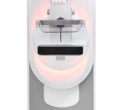
MRI-guided breast biopsy and its rapid market acceptance is a prime example of adapting technology to imaging. The MRI biopsy market is growing at 60 percent per year and is expected to produce more than 1 million scans and 200,000 biopsies by 2010 – Frost and Sullivan projects a 35 percent annual growth rate in the number of MRI-guided breast biopsy procedures from 2005 to 2010. Today, more than 8,000 vacuum-assisted MRI-guided breast biopsy procedures have been successfully completed using the ATEC.
Chris Bleck, CEO, Intact Medical Corp., Natick, MA
Most of the recent innovations focus on user benefits – quicker procedure times or fewer steps to retrieve the tissue specimen. Unfortunately, almost all biopsy systems provide the pathologist with a fragmented histology sample. As a result, many specimens do not allow the pathologist to render a definitive diagnosis. The next meaningful innovation will be minimally invasive systems that permit the user to retrieve a larger, intact tissue specimen to the pathologist for a diagnosis. The goal of these newer systems is to aid the physician in improving the positive predictive value of breast biopsies.
Future innovations will continue to be minimally invasive and provide tissue specimens that have the same quality histology as an open surgical procedure. The best cutting method to achieve this goal is radio frequency (RF), which allows the target lesion to be removed intact. This allows the pathologist to render a diagnosis from the intact target lesion and the tissue surrounding the target. This will maintain the tissue architecture similar to the tissue specimen from an open surgical excision. These systems allow for quick tissue capture, easy advancement through dense breast tissue and minimal bleeding due to the vessel sealing properties of RF devices.
The systems that allow for the quickest procedure time will be successful since they minimize the time that patients are required to be in uncomfortable positions. This also improves patient throughput for busy clinical practices and permits more procedures to be performed on a daily basis. Since patients are awake for these procedures, patient comfort and pain management are very important. Many times, this is more dependent on the anxiety level of the patient and administration of anesthesia than the type of device that is employed. It is important to use the system that has the highest likelihood to provide a definitive diagnosis. This will minimize the number of patients who require re-biopsy or an open surgical procedure.
Regarding easy-to-use plug-and-play imaging modalities (e.g., one-click switching to ultrasound, MRI or CT): It is not necessary to conduct multiple studies on the patient immediately. However, the physician may find it convenient to have all imaging studies available on the same system.
Convergence with imaging modalities and integration with 3-D CAD programs may be of benefit to the physician to have all of the information at their fingertips when making a diagnosis.
Adjustable or changeable collection mechanisms (e.g., one-click switching to graspers or pouches from vacuum filters) will be important as the procedures migrate to specimen collection systems, which preserve the intact architecture of the tissue. This system is currently available from the Intact Medical Corp. and provides multiple tissue capture options.
On automatic pain management medication delivery: Yes, pain management is important to the patient and the physician alike. Physicians would like to optimize the patient experience so patients will continue to serve as a source of referral.
On cryoablation versus laser ablation: Adoption of these methods may continue to be slow since the data on long-term outcomes compared to surgical options is sparse. The current uses of cryoablation or laser ablation are limited to treatment of benign conditions and rarely impact the treatment outcome of the patient.
Intact tissue collection will be of value since it is a quick method and provides histology specimens similar to open surgical excision biopsies. Superior specimen collection techniques are cost-effective and reimbursement is currently available under existing codes to encourage use of these techniques. RF cutting methods are very quick and can dramatically reduce the time required per procedure.
Michael Terlizzi, vice president, sales and marketing, Bard Biopsy Systems, Tempe, AZ
Like much of the device industry, the customer’s needs will drive the next generation of products that bring a solution to the problem of accurate diagnosis of a breast lesion with the least intervention.
Even at this time, with many new devices taking larger samples, a large segment of patients still have surgical biopsies. In addition, while vacuum-assisted biopsy has become the standard of care when stereotactic imaging is used, most ultrasound-guided biopsies are still acquired with a core needle, which produces a significantly smaller sample size for pathology.
The use of MRI will also increase the number of biopsies as lesions are recognized at an earlier stage.
All three of these dynamics will lead to the next generation of devices meant to respond to the problem of accurate diagnosis with the least intervention.
I believe the improvements in ultrasound technology will result in more physicians becoming comfortable using ultrasound-guided biopsies as an alternative to surgery.
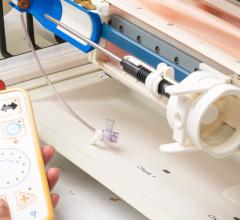
As this dynamic occurs, the need for a biopsy device that simplifies the acquisition of a lesion, leading to a more accurate diagnosis will become the target. The Bard Vacora Vacuum Assisted System is the first self-contained, vacuum assisted biopsy device that delivers a larger sample, simple operation and no significant capital investment.
I believe as we move forward, biopsy devices will continue to evolve to simplify the physician’s technique, improve diagnostic accuracy and increase patient comfort with a less invasive approach.
James Vetter, M.D., chairman and CEO, Rubicor Medical Inc., Redwood City, CA
Sometimes going forward means going back….to some very important principles. ‘Way back’ to OSB — for important reasons the predominant biopsy procedure.
These historic, important principles need not be compromised for the sake of ‘minimally invasive.’ And ‘minimally invasive’ is extremely important for many reasons — scheduling, cosmesis, healing, patient satisfaction and cost containment.
What are these principles that should not be ignored? Knowing them will make the ideal solution obvious.
1. The purpose of breast biopsy is to remove tissue for accurate diagnosis and often for therapy. This dual purpose has historically necessitated multiple steps. But in appropriate cases, Rubicor’s single step means this is not always true. There appropriately had been reluctance to switch to any kind of percutaneous biopsy that compromised accurate diagnosis and did not completely remove the entire lesion in one piece. For this reason the majority have remained open surgical.
2. Breast lesions come in a variety of types. For those that need biopsy, in the majority of cases the most desirable procedure is to remove the entire lesion whole — for benign and malignant types.
3. The single most important diagnostic predictor for malignancies is their size. When the whole intact piece is extracted, accurate measurement and verification of complete removal can readily be accomplished using examination of the borders to confirm that normal tissue surrounds the entire piece — ‘clear margins.’ Little debate exists over these critical pieces of information.
If the device needs to destroy the lesion to accomplish ‘minimally invasive,’ then the precious diagnostic tissue has been wasted — eliminates ablative techniques — laser, cryoablation, etc. If the device needs to cut the target up into multiple pieces for removal then the advantages of keeping the lesion whole have been totally lost — ‘core’ biopsy.
If I were a patient, I’d want no questions remaining after my procedure. I’d want my doctor to do the most accurate diagnostic procedure without compromise and one that would also provide the best resolution — complete removal. I would love to avoid stitches, hospitals, scars and discomfort. I’d like, where possible, to have it all done at once rather than in many stages.
If a system could accomplish all that comfortably in the office it would be the ideal alternative. If it used the most accurate guidance methods to prevent excessive tissue removal the cosmetic result would be optimized.
The ideal system is Rubicor. These devices are office-based, use state-of-the-art guidance and are compatible with off-the-shelf equipment. Rubicor’s are the only devices capable of removing entire large lesions through a small skin nick while preserving features pathologists rely on for accurate prognosis and to guide other possible follow-up therapy. ‘High tech’ in this field translates to truly meeting the needs of today’s patients and their caregivers (office-based, affordable, cosmetic, comfortable and complete, whole removal).
Doug Bradley, director of marketing, SenoRx Inc. Aliso Viejo, CA
Significant advancements are being made with medical devices and in particular breast biopsy devices as a result of technological innovation with computer software/hardware integration and miniaturization. These new technologies enable the creation of ‘smart’ devices that enable the physician to customize the procedure for each patient. For example, breast lesions can be found in difficult to access areas such as close to the chest wall or close to the surface of the skin. With EnCor, the physician has the capability to program a specific sampling pattern into the device and then the device automatically samples, rotates and transfers the targeted tissue to the collection chamber. The ability to electronically adjust for full or half samples, or dense versus soft tissue acquisition or automatic anesthetic delivery are just a few of the many options that help the physician customize the procedure for the individual needs of the patient.
With the continued rising cost of healthcare, premature capital obsolescence is a key concern to physicians, particularly in an outpatient setting. Computer-controlled hardware minimizes this concern as new features can be added through a simple in-office programming change.
The trend towards ‘closed’ biopsy systems will also continue. A ‘closed’ system collects tissue from inside the breast and then transfers the tissue to a sterile holding container minimizing procedure time and cleanup. Further enhancements to the ‘closed’ system include the ability to view the samples during the procedure as they are being harvested. Oftentimes the physician can tell by looking at the tissue if the sample is being harvested from the right location. EnCor offers this feature today. Not only can the samples be viewed during the case but the doctor has the option to remove each sample at a time or wait to remove all the samples at the end of the procedure.
Replacement collection chambers that allow real-time direct X-ray of the specimen while still in the chamber will also improve the overall efficiency of the procedure.
In terms of convenience, wireless technology holds promise. Providing a remote control unit for the doctor that communicates with the main console offers the advantage of portability in addition to all the features that the main console provides.
Integration of imaging modalities with the biopsy unit will further simplify the biopsy procedure. Smart/flexible software will capture the lesion coordinates from the imaging device and transfer the coordinates directly into the biopsy device, which will then automatically target and retrieve the desired sample.
On the therapeutic side, cryoablation or laser ablation holds long-term promise if it can be documented that every cancer cell has, in fact, been destroyed. Most likely the FDA will require five-year studies, which could delay these new technologies for many years to come.
Minimally invasive therapy may also involve devices that can customize the cavity site to better match the brachytherapy balloon. Creating a more dimensionally ideal cavity could enhance radiation therapy.

 February 18, 2026