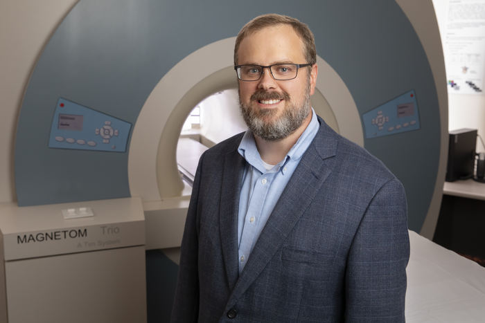
Brad Sutton, a professor of bioengineering at the University of Illinois Urbana-Champaign and the technical director of the Biomedical Imaging Center at the Beckman Institute for Advanced Science and Technology. Image courtesy of the Beckman Institute Communications Office.
March 16, 2023, marks 50 years since Paul Lauterbur published his seminal Nature paper establishing zeugmatography — now familiar to most as magnetic resonance imaging or simply MRI — as a viable way to visualize objects with a magnetic field and radiofrequency signals.
A faculty member at Stony Brook University in New York at the time of the discovery, Lauterbur was recruited to the University of Illinois Urbana-Champaign in the 1980s and won the 2003 Nobel Prize in Physiology or Medicine for developing MRI along with British physicist Sir Peter Mansfield.
Lauterbur’s first human MRI scanner is preserved in the Illinois MRI Exhibit at the Beckman Institute for Advanced Science and Technology, where cutting-edge advancements in medical imaging include:
- Advanced bloodflow imaging in the brain to better understand tinnitus.
- The first MRI atlas of a bearded dragon brain.
- Efforts to democratize neuroimaging data in diverse communities.
Most recently, researchers have unlocked the ability to conduct scans in real time and see the physical mechanics of activities like speaking, singing, and swallowing. They have also developed techniques to use MRI to visualize genetic expression in the brain when learning.
Brad Sutton, a professor of bioengineering at the University of Illinois Urbana-Champaign and the technical director of the Biomedical Imaging Center at the Beckman Institute, shares his thoughts on the momentous anniversary. He can be reached via email at [email protected].
How has MRI technology changed the scope of medical research in the last 50 years?
MRI has become one of the most important tools for doctors to see inside the body to understand what is happening in disease. MRI shows soft tissues like the brain, the heart, and other muscles and organs. It provides several ways to view the status of the tissue, such as looking at the shape, changes to the structure, blood flow, and inflammation. Being able to see inside the body quickly and clearly has led to advanced treatments and longer, healthier lives. MRI is a flexible imaging technique, and many physicians, scientists, and engineers continue to develop new ways to see disease earlier, enabling more effective treatments.
The MRI scanners themselves continue to improve. One way that the scanners have changed is the magnetic field strength. This is measured in Teslas as the unit — the earth’s magnetic field is approximately 0.00005 Tesla. Paul Lauterbur’s first human MRI magnet was 0.09 Tesla, or about 2,000 times the Earth’s magnetic field. This enabled him to see structures in the body, but grainy and at low resolutions. Modern clinical MRI systems are 3 Tesla. Recently, the University of Illinois Urbana-Champaign and Carle Hospital jointly purchased an MRI magnet that is 7 Tesla. With this higher magnetic field strength, 75 times stronger than Lauterbur’s initial magnet, we can localize function in the brain down to about 0.5 millimeters, clearly and with excellent contrast.
How does advancing MRI technology help protect human health, especially in an increasingly aging population?
New imaging technologies using MRI allow us to see how the body changes with age and disease, and how the body responds to interventions. For example, we can see how the brain changes as we get older. It is not just that important parts of the brain decrease in size; the way in which different parts of the brain communicate with each other also changes. This leads to less efficient processing of information and can lead to disruptions in things like decision-making.
As the population ages, we need effective interventions that will allow us to maintain our brain function late into life. MRI is helping with this too, enabling clinical trials on drugs that impact the brain, but also on non-pharmaceutical interventions like aerobic exercise, yoga, and brain-specific training.
What can we expect or hope for from the next 50 years of MRI research?
In the next few years, we will see new MRI systems with even higher magnetic fields, providing even higher spatial resolution images of the body and brain.
At the same time, we are also seeing new MRI systems that are small and portable, which can be placed in the doctor’s office for easy access. We will see systems that integrate information across all patients to better understand what we are seeing in the image and what it means for the health of the patient. We will also see new information when looking at the images, with new techniques leading to images where the signal intensity in the image represents quantitative information about the status of the tissue, including concentrations of key molecules in each pixel of the image, mechanical and electrical properties of the tissues, information about how the brain is performing its activities including changes to the tissue structure and genetic expression, and the systems will produce actionable 3D visualizations of the person in the scanner so that a doctor can perform virtual interventions and virtual surgeries to see the best way to treat the patient.
Given the pace of development from when Paul Lauterbur imaged his first living sample (a clam) until now, I am certain that we will see these developments before another 50 years.
For more information: https://beckman.illinois.edu/

 July 20, 2026


