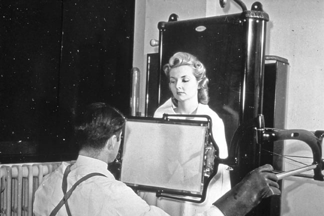
Shown is a male technician taking an X-ray of a female patient, circa 1940. This image was used to demonstrate the myth about exposure to radiation during the X-ray procedure. Source: National Cancer Institute, National Institutes of Health.
The group known today as the Society for Imaging Informatics in Medicine (SIIM) had its origins in 1980, when a small group of individuals who saw that medical imaging was on the verge of undergoing a revolutionary change met and formed the Radiology Information System Consortium (RISC). It was comprised of academic and private hospitals for the purpose of developing a radiology information system (RIS) with a commercial partner and making it available to member institutions and others.
In 1989, the Society for Computer Applications in Radiology (SCAR) was founded as an organization under the RISC banner. SCAR provided a structure to include individual members, and RISC’s biannual meeting on computer applications was transferred to SCAR in 1990. In 1996, RISC and SCAR merged under the SCAR name to include individual, corporate and institutional members.
In 2006, the Society changed its name to the Society for Imaging Informatics in Medicine to better describe the diversity of its constituency, to expand research and activities into the entire field of imaging informatics, to include all of the imaging sciences and to embrace the dynamic changes in the healthcare environment.
A Rich Historical Archive
SIIM documented and archived a large amount of historical information about its organization and segments of the medical imaging industry, which can be found on its website, www.siim.org. These include videotaped and transcribed reminiscences from a founding member, Joseph N. Gitlin, DPH, FSIIM, associate professor of radiology, Johns Hopkins Medical Institutions, which provide an inside look at important milestones, as Gitlin’s career often was interwoven with developments in the industry.
Gitlin graduated from the University of Pennsylvania and early in his career worked for the U.S. Public Health Service (PHS). While there, he earned a master’s of public health degree at Johns Hopkins. A few excerpts of his transcript are presented below, based on the interview conducted by Steven Horii, M.D., FSIIM, University of Pennsylvania Health System and chairman of SIIM’s History Committee at the time of the taping.
Fighting Tuberculosis With X-Rays
“When I was accepted in the Public Health Service in 1950, my first assignment was with the Division of Tuberculosis, when a national screening program was underway to make an attempt to eliminate tuberculosis in the United States. The technique that was developed included a photofluorographic unit that recorded chest X-ray examination images on 70 mm square film…The PHS had a laboratory…known as ‘Murphy’s Lab.’ That’s where the new photofluorographic X-ray unit was tested under the direction of Dr. Russell Morgan, who was active in the development of the test procedures. At the time, Dr. Morgan was the chairman of the Department of Radiology at Johns Hopkins and a consultant to the PHS.
“The work resulted in 60 mobile units being built, each of which was equipped with the photofluorographic X-ray equipment. Over the next three years of operation, there were 2 million chest X-ray examinations done throughout the country. It was a free program, highly publicized and very well attended. A large number of cases of previously unknown tuberculosis were found that were treatable because isoniazid was available. Instead of sending patients with active disease off to sanitaria, the treatment was usually performed at home.
“During this assignment, I participated in designing a punch-card system that recorded the fact that an individual had participated in the screening program and provided names and addresses, so that if a 70 mm reading was positive, followup could be performed. The punch cards also were used to produce program statistics. A professor of statistics at the University of California analyzed the data, and we noticed that there were relatively high positive rates of readings by the young radiologists who were interpreting the 70 mm films.
“As a result, we got permission and funding to change the single reading of the 70 mm films to independent double reading. That pretty much solved the problem of ‘over-reading,’ and it greatly reduced the follow-up costs, because every positive reading on the small films resulted in the individual going to a hospital or clinic and having a 14-by-17 film exam done to confirm the positive screening result.
“The only thing digital we knew about in those days was that you used your hands and your fingers to punch the holes in the cards…A special reader was developed to hold the 70 mm films that were on a roll of 500 and magnify them to get a better view without changing the image proportions. But there was a tendency, because of the screening program and single reading, that if the reader saw anything that was abnormal it was recorded as positive and that resulted in calling the patient back for the 14-by-17.”
Early Radiation Safety Concerns
Another change that affected the industry as well as Gitlin’s career came about in 1957, when President Dwight D. Eisenhower issued an executive order that removed the Health and Safety regulatory authority from the Atomic Energy Commission. The commission was responsible for promoting the use of radionuclides and radiation activities, including energy production. As Gitlin noted, “It spent 90 percent of its time promoting it rather than giving priority to health and safety.
“At that time, we were testing weapons in the atmosphere and there was a great deal of concern about fallout and dietary ingestion that contained a variety of radionuclides. When President Eisenhower transferred that responsibility to the PHS, that, in effect, became the beginning of the organization of the Center for Radiological Health. I was assigned to the new Radiological Health Laboratory in Rockville, Md….We set up a regular monitoring program with a PHS ‘Air Force’ made up of four planes based in Las Vegas and many monitoring devices that collected an enormous amount of data.
“Initially, we used punch cards to record the data, but it was clear that better data processing methods were needed. I was successful in arranging with IBM to install its model 1620 computer, which in effect was the first solid-state, mid-sized unit available. It was a scientific high-speed unit that we installed in the Rockville laboratory to process all the data that we collected from 1960 to 1963, the period when atmospheric testing of weapons was still conducted.
“The data collection and processing effort placed special emphasis on strontium 90, which was of great public health concern because of infants and children drinking milk and possibly ingesting strontium 90. The program had a lot of important publicity attached to it and people followed the weekly reports very, very carefully. The atmospheric weapons testing ban was signed by President Kennedy in 1963, several months before he was assassinated. The ban halted the testing by the U.S. and other countries that had signed the treaty. The monitoring program continued for almost two years afterward just to be sure nothing else was happening.
“The design included household interview surveys from a representative sample of the population, in which the woman of the house usually provided information about the food consumed during the previous week…The program worked quite well and we saw a downward change in the radioactive measurements over the four-year period of the food collection program. This clearly showed that the atmospheric weapons testing ban had, in fact, achieved its goal.
An Early Start to Measuring Exposure
“The work that we did in household interview surveying gave rise to the idea that we could produce estimates of population exposure to X-ray examinations by asking people in households if they had any X-ray exams, including dental, during a specified period of time. A serious statistical problem was associated with picking the time period. If people were asked about X-ray examinations in the past year, the responses provided relatively large numbers of procedures, but because of the memory factor, there were many false positives and negatives. If the time period was the past month, the result was relatively few exams with few false positives and negatives.
“We compromised by choosing a three-month time period that was tested in a pilot study of the protocol in Berks County and Reading, Penn…The county health officer there was an old friend of mine, and he encouraged residents to participate in the household interview and the follow-up phases of the study.
“In addition to showing that residents were able to report that they had medical and dental X-ray examinations, we developed an X-ray film pack to obtain radiation exposure data on each exam reported in the household interview phase. Patients provided permission for a follow-up visit by a PHS officer to the clinic, hospital or physician who did the exam to verify the report and perform an exact replica of the procedure using the film pack as the object. After the film pack was exposed, it was sent back to Rockville, where it was processed, and estimated exposure to various organs were recorded.
“These data were translated later into dose estimates using a computer program at Johns Hopkins, where I worked with Dr. Russell Morgan and a staff of experts. The results of the X-ray exposure study were very well received by physicians, scientists and public health officers. The findings facilitated improvements in the training of technicians and the education of interpreting physicians.
“We found that one of the most important problems with X-ray examinations in those days was that the size of the X-ray beam used was often far larger than the organs of interest and was exposing much more of the patient’s anatomy than was necessary. This led to the development of beam limiting devices by the manufacturers, which was installed on all types of X-ray machines.
“As I mentioned earlier, Dr. Morgan and the computer staff at Hopkins were very cooperative in writing the programs and developing the conversion tables. When that was finished, we tested the validity of the organ dose tables in cooperation with Dr. Richard Chamberlain at the University of Pennsylvania to estimate dose from selected X-ray exams in certain organs. When it was feasible to measure dose during examinations with the patient’s permission, we compared the measurements with the estimates produced by conversion tables developed at Hopkins.
“When we were satisfied that the computer programs worked well, I began to think about what should be done next. The staff in Rockville wanted to write two papers related to the 1964 national study and I agreed. We produced a report on X-ray examinations during pregnancy as a subset of what we learned about the general population in ’64, and a report on the dose estimates based upon the ’64 exposure data and the conversion tables.”
The Era of Data Processing
“At Hopkins, we began to recognize the potential for using data processing systems in radiology in the day-to-day operations. Dr. John Dorst, director of pediatric radiology, had set up a punch-card system to record each examination that was done in his section to expedite the searching of the film library for specific exams…When I learned about the use of the punch-card system, with Dr. Morgan’s approval, we began to record data on a punch card for every X-ray exam that was done in the department during 1966.
“An analysis of the exam data on the punch cards helped settle an argument between the comptroller’s office of the hospital and the radiology department regarding the number of exams performed. The comptroller’s office always seemed to underestimate the income derived from radiology, which was a factor in determining what the budget for next year would be. And so, in his usual fashion, Dr. Morgan said, ‘We ought to have a verification process.’
“He asked me to participate in expanding the initial punch-card system to cover the entire department, and as it turned out, we had 6 percent more examinations based on the punch-card system than the comptroller had from the paper process of several layers to produce financial reports. Dr. Morgan was very pleased with the additional dollars in next year’s budget, and we began looking at other areas of radiology that might benefit from computer applications.
“A preliminary study of departmental activities was conducted at Hopkins. It included radiologists at Hopkins and several members of the PHS radiological health program in Rockville. Data were collected on each of the recurring activities at Hopkins, and similar baseline studies were conducted at other institutions to determine where computers could help radiology departments become more efficient. The activities included scheduling patients and exam rooms, film library management, the reporting of interpretations of films that often took three to four days when they were dictated and transcribed, and support for teaching and research activities from records on interesting cases.
“In almost every instance, the baseline studies at Hopkins and other institutions that used similar protocols identified the same kinds of problems and suggestions that computers could help in these areas, and this was before we could actually do it, but the concepts were defined.”
To read more of the transcript or view the video interview of Joseph Gitlin and Steven Horii, coordinated by the SIIM History Committee in August 2008, visit www.siim.org.
SIDEBAR:
X-Rays and Shoes
The SIIM History Committee’s video interview with Joseph Gitlin included a sidebar on one of the more prevalent uses for X-rays about 50 years ago — to determine shoe sizes. Following are Gatlin’s memories about it.
“A sidebar to the X-ray exposure study concerns fluoroscopic shoe fitting machines. Some of you may not remember when your mother took you to the shoe store in the ’30s, ’40s, ’50s; kids enjoyed trying on their new shoes, walking over to the shoe fluoroscopy unit, putting their feet in and watching their toes wiggle. Mothers were interested in getting the right size shoes for the kids with space so they could last another six months.
“The position of the fluoro unit could produce gonadal exposure as well as foot images. In 1958, we banned shoe fluoroscopy in the United States. But in 1964, when we conducted the national study, we soon had people in the household survey report X-ray exams as being done at a shoe store that was not aware of the ban.
“The Surgeon General wrote a letter to the state health officers indicating that any shoe store that had a fluoro unit but collaborated by providing data in the study would not be prosecuted if it stopped using the unit. One day in 1964, I received a call from the regional representative in North Dakota, saying, ‘We have a shoe store operating a shoe fluoroscopy unit, what should we do?’
“And I said, ‘Well, read the letter from the Surgeon General and tell the owner that he is exempt from prosecution if he stops using it. And if the owner of the store offered to donate the unit to the Rad Health Museum in Rockville, we would pay the freight.’ He made the offer, and the last known shoe fluoroscopy unit in the country is on display.”
Read the related article "The Eclectic History of Medical Imaging."

 July 20, 2026


