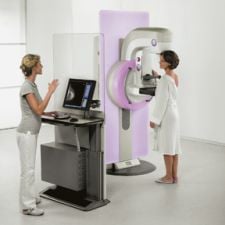
Photo courtesy of Siemens Medical Solutions
Less than three years ago, a small number of outpatient imaging centers were taking the wraps off machines to perform mammograms most insurers likely wouldn’t cover.
But today, thanks in large measure to a landmark nationwide study confirming its efficacy as a screening tool superior to film-based systems for a large segment of the female population, digital mammography is now poised to overtake film as a dominant breast screening modality – particularly in the outpatient arena.
Demand for digital systems is so brisk that market researchers can hardly keep up with the pace of technological innovations as manufacturers try to differentiate themselves with new digital detectors, high-tech tubes and sophisticated software for enhancing, manipulating, transporting and archiving data and images.
Boon Times for FFDM
Barely a smattering of hospitals and outpatient diagnostic imaging centers were toying with full-field digital mammography (FFDM) systems when the National Cancer Institute (NCI) released results of a four-year, multicenter 50,000-patient clinical trial whose aim was to determine whether it is equal or superior to standard film mammography.
Objectives of the $26.3 million Digital Mammographic Imaging Screening Trial (DMIST), the largest federally-funded clinical trial on medical imaging and conducted by the American College of Radiology Imaging Network (ACRIN), were to determine whether FFDM was better than film mammograms at detecting abnormalities and differentiating between benign and malignant tumors, and, perhaps, the 15-20 percent of breast cancers that now go undetected in film mammography.
The results of the trial were a boon for imaging companies. Researchers, in fact, concluded there was virtually no difference between X-ray film and digital mammography among women in the general population. Moreover, digital mammography was shown to be superior to film for women who are pre- and perimenopausal, under age 50, and/or with dense breasts.
“Digital mammography is getting much closer than ever before in replacing film as the gold standard of care,” said Andrew Vandergrift, national marketing manager, Women’s Healthcare Imaging Systems, for Stamford, CT-based FUJIFILM Medical Systems USA. “The market was really hanging on the results of the ACRIN study. So much was hinging on those results.”
The door has swung wide open, in fact.
“We’re seeing an accelerated growth in demand for digital mammography. This has placed additional pressures on manufacturers to deliver digital systems,” said David Caumartin, global general manager, Mammography, for Wauwatosa, WI-based GE Healthcare Clinical Systems. “The ACRIN study showed the superiority of digital mammography in spotting tumors in dense breasts. This has been the hard piece for radiologists.”
“The market is speaking with their pocketbooks,” Vandergrift told Outpatient Care Technology, adding that demand for FFDM systems in the outpatient arena has been “dramatic.” Before the ACRIN study, only about four percent of all centers were doing digital mammography. Today, about 30 percent of outpatient imaging centers are using FFDM, Vandergrift added, and the market is growing at about 80 percent per year.
Vandergrift’s figures are consistent with those from the FDA, which reported that there are currently 3,945 facilities with FFDM units. The FDA MQSA Facility Scorecard, in fact, noted that 30 percent of the 8,837 mammography sites in the country have at least one digital mammography system.
So swift is the buying, that it is far eclipsing sales of conventional X-ray systems, said Jim Culley, Ph.D., director, Strategic Projects, for Danbury, CT-based Hologic Inc. “NEMA (the National Electrical Manufacturers Association) reports that less than 8 percent of the mammography system sales in the U.S. for the 12 months ending in August 2007 were screen-film systems,” he said. “Everyone else bought digital. The FDA MQSA monthly scorecard shows that 30 percent of the 8,837 MQSA sites in the U.S. now have at least one digital system -- up 84 percent in just 12 months,” Culley told OPCT.
For outpatient imaging centers, digital mammography couldn’t come at a better time, considering the efficiencies it allows an industry strapped by higher workloads and a shrinking pool of radiologists. “The number of imaging centers is actually decreasing now because of consolidation, so it’s becoming more and more important for machines to be easy to use and to allow techs to do centralized reading,” said Vandergrift.
What’s Fueling the Growth?
A bevy of technical capabilities, more educated clinicians and consumers and considerable revenue potential all are driving demand for digital mammography among the outpatient and acute care segments.
Digital mammography units today have a host of benefits over their film-based counterparts, including:
• Improved image quality;
• Ability of clinicians to manipulate images;
• Reduced radiation exposure to patients;
• Superior dose and contrast performance;
• Digital archiving and display;
• Seamless connection to information systems;
• Higher clinician confidence;
• Reduced number of callbacks; and
• Reduced exam time, increased efficiency and throughput.
In addition, more educated consumers and buyers are fueling growth and acceptance. Prompted by swelling customer demands, facilities like Jane Phillips Imaging Center, a Bartlesville, OK-based facility, are rapidly converting to digital. Jane Phillips plans to triple its digital mammography service line across three centers by spring 2008 since upgrading all of its existing film mammography equipment to the latest in digital technology.
There’s also more revenue to be had in digital mammography. Transitioning to digital systems reportedly enabled one large Midwest breast imaging center to perform an additional 10,000 procedures per year, and generate more than $1 million a year additional revenues for surgery and radiation therapy.
And for the first time, the reimbursement climate favors digital mammography.
According to Vandergrift, digital systems also have much higher reimbursement levels than film today, 50 percent or higher in many states. Still, reimbursement can vary from state to state, as one recent published study found. Some insurers, meanwhile, are taking a wait-and-see approach. For example, an official with Health First, the largest insurer in Brevard County, Florida, told Florida Today, a local newspaper, that the company hasn’t completely embraced the ACRIN study findings, and requires its members to obtain permission before getting a digital mammogram.
Film, CR Have Staying Power
Meanwhile, it remains unclear whether conventional film mammography will ever be completely supplanted by digital since the ACRIN study limited digital’s superiority to a smaller sub-class of female patients.
“Film is still in high demand, especially in the outpatient arena,” said Fred Pilgrom, technical product manager, special products-woman's health, for Malvern, PA-based Siemens Healthcare, who added a significant number of the company’s outpatient customers are still buying and using film.
A study released in January 2008 and reported on in the Annals of Internal Medicine, hinted that centers may not be prudent to get rid of their film-based systems and go all digital since such systems are not cost-effective for screening patients outside of the class of women identified by the ACRIN trial. “Relative to film mammography, screening for breast cancer by using all-digital mammography is not cost effective,” researchers noted. “Age-targeted screening with digital mammography seems cost effective, whereas density-targeted screening strategies are more costly and of uncertain value, particularly among women age 65 years or older.”
Moreover, the future of computed radiography as a breast-screening tool doesn’t seem to be in jeopardy, either, in spite of it once being seen as a potentially widely used modality for mammography. That said, some observers say CR still falls short of the image quality offered by FFDM.
“CR hasn’t really taken off for several reasons,” said Caumartin.” For one, the workflow is about equal to that of film and its ability to detect cancer is not that much better than film.” Moreover, he added, computer-aided detection (CAD) systems, known to increase diagnostic confidence, “don’t work that well with CR.”
However, many still view CR as a viable tool, if only as a bridge for many centers wanting to make the digital leap. Just ask officials with Rochester, NY-based Carestream Health. Carestream has a new platform for its CR-based digital mammography “solutions” and a fully featured digitizer that converts film-based mammograms for efficient digital image review.
“While initial digital mammography systems had small fields of view that made them suitable for diagnostic work only, the introduction of CR and DR systems enables the technology to be used for screening exams as well,” said Stephen Archer, director of Worldwide Marketing, Mammography Solutions, for Carestream Health. Archer noted buyers should recognize that “digital mammography comprises more than just the capture modality. It’s a full solution that incorporates image processing, interpretation, display, sharing and storage.
“We do believe there is significant room for improvement in both digital and film-screen technologies and we expect continuing advances in accuracy and clinical effectiveness in the future,” Archer added.
Companies such as FUJIFILM also believe strongly in CR, incorporating the technology in its line of Fuji CR mammography (FCRm) units. “CR may speed the transition to digital mammography because it doesn’t require investment in entirely new system,” Gillian Newstead, M.D., director of Breast Imaging at the University of Chicago, noted in a video posted on Fuji’s Web site. “It also can be used with existing equipment, and allows digital storage and image manipulation.”
Still, as alluring as digital mammography is for many outpatient centers seeking to attract more patients and more revenue, it’s no easy task converting from a film world.
As GE Healthcare Clinical Systems noted in a white paper posted on the company Web site, “converting to digital mammography means more than replacing one piece of equipment with another. It means building a complete imaging workflow that includes image capture, distribution, review and archiving.
“Mammography remains a lower-margin offering, unforgiving of waste and inefficiency. Facilities that approach digital conversion as ‘just another piece of equipment’ often experience failure,” the company added. For example, one breast imaging center initially failed in its conversion to digital because it did not redesign its workflow to complement the technology. Instead, the center kept the same exam time lengths that they had used in the analog environment.
Successful film-to-digital conversions also entail helping clinicians compare apples to apples, or in this case, prior films with new digital images from the same patient. “Radiologists today are hindered because they are being forced to compare a patient’s earlier X-ray films with digital images taken today,” said Vandergrift.
To that end, most manufacturers, including FUJIFILM and Carestream, incorporate capabilities into their systems that attempt to ease the ability to compare digitized films with digital images.
Carestream, for example, offers a digitizer that builds on the company’s goal to help mammography facilities transition to digital imaging by converting prior film-based diagnostic and screening mammograms to a DICOM-based digital imaging format so they can be viewed in conjunction with digital mammography exams on a diagnostic workstation. This eliminates the need for radiologists to review prior exams on a film box while digital exams are reviewed in soft copy.
Hologic, which enjoys a dominant presence in both the digital and film-based markets, offers its own solution called “R2 DigitalNow,” which it dubs “the bridge to a digital environment.” DigitalNow also provides the capability to scan films for soft-copy reading, archive images for future use and get CAD results.
Brisk Innovation
Among manufacturers, the pace of innovation is eclipsed only by unit sales.
Those with whom OPCT spoke were nearly unanimous in stating that a great deal of current innovation resides in improving image acquisition and processing.
“Software is where a great deal of innovation is occurring right now,” said Vandergrift. “There’s a lot of exciting work going on with image processing algorithms, information management and data compression.” Pilgrom agreed. “A lot of effort right now is in improving image resolution and quality,” he said.
Here’s a look at some of the key features of digital systems.
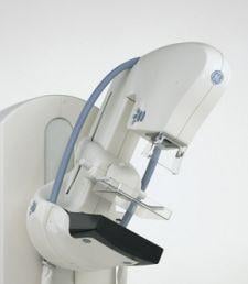
• Digital detectors – Perhaps the most important component of any FFDM system, detectors are image acquisition devices. And today, the operative term is “full field,” which refers to the ability of displaying breast images as large high-resolution images, typically 24 x 30 cm – the size of most large screen-film cassettes. Detectors are essentially of two main varieties: indirect and direct. Indirect versions use a scintillator layer to convert X-rays into light, while a photodiode layer composed of tiny charge-coupled devices converts the light into digital data. Direct detectors bypass this step and convert the X-rays directly into electrical charges and data. Manufacturers that use such detectors, including Hologic, claim they produce a superior image because conversion of X-rays into light degrades the data. Because a third or more of women are classified as having large breasts, today’s digital detectors must be capable of capturing the entire breast in one shot. GE Healthcare claims that its Senographe Essential’s detector is designed specifically for mammography and offers a 24 x 31 cm size – the largest field of view in the industry. Caumartin said GE Healthcare invested $135 million in a dedicated mammography detector manufacturing plant in New York State, “a huge commitment on our part.”
• Image quality – Any discerning buyer of a digital system has done their homework when it comes to pixels, which physicists will debate endlessly over. Nonetheless, with microcalcifications as small as 100 to 200 microns, detector pixels can be a critical buying decision. (Significant advancements in X-ray film allow conventional systems to spot microcalcifications far below 45-microns in size.) Experts say when it comes to detectors, the smaller the pixel, the higher the resolution. FUJIFILM’s ClearView 1M and CSM systems boast 50-micron pixel sampling, for example, and are capable of reading both sides of the plate, Vandergrift says. Some users, meanwhile, say higher pixels don’t necessarily mean lower image quality. Eastern Radiologists Inc. Breast Imaging Center, for example, told GE Healthcare that the images from the 100-micron detector on their Senographe Essential were superior to a 50-micron detector they previously used.
• Advanced image-processing techniques – The secret’s in the software, and most manufacturers offer various kinds of advanced tools to manipulate digital images. Fuji says its “dynamic range control” improves the visibility of both dense and soft tissue by amplifying or reducing image density and contrast. Carestream’s “Mammography Digitizing System” has automatic processing capability that allows up to 50 film images to be scanned without operator intervention. Hologic incorporates Advanced Image Enhancement Inc.’s (AIE) software for examining “regions of interest” in its SeleniaT digital mammography system. The software, based on AIE’s expertise in locating and detecting undersea mines, is said to improve the conspicuity and detail of calcifications.
• Multimodality capability – A common feature with digital systems, the multimodality feature is an essential one in centers that offer a variety of imaging services. “SecurView diagnostic workstations proved consistent display of images from different vendors, in conformance with the IHE Mammography Image Profile,” said Culley. “Prior images are automatically adjusted, displayed and printed the same size as images of the current patient evaluation, regardless of differences in detector size, detector resolution or paddle size. And an integrated LCD color display lets you perform side-by-side review of mammography images with DICOM images from other breast imaging modalities, such as ultrasound and MRI.”
• CAD interface – CAD technology allows clinicians to distinguish cancers from other breast abnormalities. Most manufacturers incorporate CAD capability. GE Healthcare and iCAD, for example, announced an agreement at RSNA 2007 to develop a customized version of SecondLook Digital for GE Healthcare’s Senographe and Seno Advantage family of systems. GE said this “next generation” CAD system improves sensitivity to breast cancer imaging, enhancing usability and workflow as well as providing diagnostic support tools. Hologic says its “R2 CAD” offering gives users the most sophisticated pattern recognition software to help find cancers with greater certainty at an earlier stage.
• Patient comfort – This is a significant concern and an even bigger marketing point for most imaging centers, and manufacturers have responded with a variety of features to minimize pain and anxiety, as well as exam time, for their patients. Siemens’ Mammomat Inspiration, scheduled for release in late 2008, features “one-click-to-image” functionality, a much faster processing capability, as well as a special “MoodLight” mode that uses LED panels that glow in different colors. A 2007 addition to the Hologic line includes the MammoPad breast cushion, whose grip-like surface holds breast tissue in place to ensure optimal positioning.

 June 02, 2026


