The last couple of years in the United States and Europe have seen an increased focus on the cumulative patient dose received from a wide variety of X-ray devices including classical X-ray, computed tomography (CT), interventional radiology and mammography. The deleterious effects of radiation dose have received widespread coverage, and the partial answer has been a movement towards non-ionizing diagnostic imaging equipment, such as magnetic resonance imaging (MRI) and ultrasound.
The increased use of ultrasound both in diagnostic and guided applications, as well as in specific venues such as the emergency room, is well documented and continues to rise. This rapid increase is expected to continue as the newer ultrasound units, especially hand-carried and hand-held units, continue to improve and as new and better software applications for noise reduction and image enhancements become available.
The relatively low-energy acoustic waves used in ultrasound, which make for a relatively safe diagnostic imaging environment, lead to difficulties penetrating thick layers of human tissue. With approximately two-thirds of the U.S. populations estimated to be obese and other estimates showing that more than 50 percent of all abdominal scans are of technically difficult patients (typically with a BMI in excess of 30), this problem dominates the diagnostic ultrasound market in both the United States and Europe. Also, various sources estimate the average exam of a difficult patient takes 20-40 percent longer – if it can even be carried out.
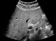
The liver and other key abdominal organs can lie 4 to 5 centimeters below the surface in difficult patients. This presents great difficulties in achieving good diagnostic quality ultrasounds and places more physical strain on sonographers and radiologists. Radiologists must use a lower frequency probe to penetrate the tissue, which leads to a concomitant loss of resolution. In cases where the low-frequency probe fails, the radiologist must resort either to a more expensive and time-consuming MRI exam or a dose-producing CT exam.
The ultrasound industry has recognized this challenge and developed a number of technologies both in software and hardware to address it. Leading probe manufacturers have attacked the problem via advanced design and materials. Software suppliers have also developed targeted approaches to penetrate the deep tissue. For example, ContextVision’s newest ultrasound image enhancement product includes a directed, focused image enhancement mode. This mode allows the user to enhance deep-lying organs without over-effecting mid- and near-field structures. When combined with the newest probe technology, this image enhancement technology will alleviate some of the issues associated with ultrasound examinations of technically difficult patients.
Donald Barry is director of commercial development for ContextVision, which is an independent developer of medical imaging enhancement, analysis and processing technologies, serving leading OEMs and distributors for more than 25 years.

 June 24, 2026


