Photo courtesy of Siemens Medical Solutions
We are living in the dawn of molecular medicine, and biomarkers represent the technology that is opening the doors to an unprecedented level of personalized medicine.
Biomarkers are facilitating a paradigm shift in the diagnosis, treatment and follow-up of patients, along with more efficient drug development. This has prompted industry, nonprofit and governmental organizations to join forces to test new biomarkers, using a variety of imaging techniques on magnetic resonance (MR) and positron emission tomography/computed tomography (PET/CT) , in particular, to accelerate disease-specific research and advance personalized medicine.
Molecular medicine gets personal
“Molecular medicine is heralding a new era in diagnostic capabilities that could change the lives of millions of Americans,” said Michael Reitermann, president, Molecular Imaging Division, Siemens Medical Solutions. “Advancing this field brings with it the promise of personalized therapeutics, which would not only improve the efficiency of healthcare, but most importantly, would also improve the quality of healthcare for patients.”
Created to have great molecular specificity, once injected into a patient, biomarkers bind to targeted, diseased cells or tissues to allow for the detection of those abnormal cells and tissues with imaging. What is so exciting about this technology is the information it provides to the medical provider. Imaging biomarkers can provide early warning signs of disease states, such as Alzheimer’s, Parkinson’s and breast cancer, lung disease and lymphoma.
Siemens Medical Solutions, GE Healthcare and Philips Medical Systems have all bought or partnered with pharmaceutical companies to develop biomarkers. The goal is for biomarkers to usher in truly personalized medicine, where a patient is genetically tested and screened for predispositions to many different diseases and then periodically screened throughout his or her life to detect the disease in its earliest stages.
“Imaging biomarkers have increasingly become an essential tool not only for the noninvasive diagnosis of disease such as cancer or cardiovascular disease but also for the development of new therapeutic drugs by the pharma and biotech industry,” said Hartmuth Kolb, Ph.D., vice president, Siemens Medical Solutions MI Biomarker Research.
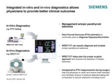
This philosophy prompted the establishment of the Siemens Medical Solutions Molecular Imaging (MI) Biomarker Research facility. The lab is dedicated solely to the discovery and development of new imaging biomarkers to spur the growth of in-vivo molecular diagnostics and is part of the company’s strategy to become a first full-service diagnostics company, integrating in- vivo and in-vitro imaging diagnostics capabilities. In collaboration with academia and industry, Siemens is developing at the site molecular imaging tracers for cell proliferation (imaging of aggressive tumors), hypoxia (to aid the planning of radiation therapy), and angiogenesis (to aid chemotherapy). Future research will branch out into neurology and cardiovascular disease areas.
Siemens also recently launched Siemens Medical Solutions Diagnostics as the in-vitro complement to the portfolio on the heels of acquisitions of Bayer Diagnostics and Los Angeles-based Diagnostic Products Corp.
Similarly, the merger of GE Medical Systems and Amersham plc was created to develop new in-vivo markers and the imaging equipment needed to produce them. The new division combines skills ranging from basic life sciences and proteomics to chemistry and engineering, with physicists, biologists, radiochemists, engineers and physicians working together on molecular imaging from all over the world.
Targeting Alzheimer’s
disease
Over the last few years, researchers have identified deposits of beta-amyloid protein in the brain as an indicator of Alzheimer’s disease (AD) prior to signs of memory loss or preclinical AD. Researchers are investigating potential
biomarkers for this debilitating disease, such as proinflammatory cytokines, as well as using MR to analyze anterior cingulate size and thickness to distinguish normal from mild AD.
Due to AD’s link to increased brain deposition of amyloid-beta (Abeta) peptides in senile plaques (SPs), recent therapeutic efforts have focused on inhibiting the production or enhancing the clearance of Abeta in brain. Investigators have developed a radioligand, [(125)I]TZDM, which binds Abeta fibrils with high affinity and labels amyloid plaques in vivo.
There is also the putrescine-gadolinium-amyloid-beta peptide probe, which targets beta-amyloid plaques and is detectable by magnetic resonance imaging (MRI) because of contrast imparted by gadolinium labeling.
Recently, Bayer Schering Pharma and Avid Radiopharmaceutical announced that Bayer Schering Pharma had exercised its right to license Avid’s 18F-AV1/ZK compound, which is a molecular imaging agent that specifically targets amyloid plaques in the brain. They hope that this imaging agent, when used with PET imaging, may lead to earlier and more accurate diagnosis of Alzheimer’s.
This personalized approach to medicine represents a shift in medical care as it focuses on patients according to their genetic make-up and the ability to perform very early diagnosis and treatment. Advances in managing AD are extremely important as the disease currently affects an estimated five million Americans, and by the year 2050, is expected to affect up to 16 million Americans.
Imaging’s critical role
Seeing is believing, which makes it no surprise that medical imaging is the technology that is unraveling the mysteries behind cellular activity, gauging therapeutic response in patients and supporting drug development. Two modalities that are particularly useful with biomarkers are MR and (Fluorodeoxyglucose) FDG-PET.
Some of the techniques used on MR to analyze biomarkers involve dynamic contrast-enhanced MRI (DCE-MRI), dynamic susceptibility change MRI (DSC-MRI), diffusion-weighted MRI, diffusion tensor imaging (DTI), MR spectroscopy (MRS) and blood-oxygen level dependent (BOLD) MRI.
Researchers are leveraging both DCE-MRI and DSC-MRI to evaluate the effects of antiangiogenic/antivascular therapy on microvascular systems. While measuring cell volume and density changes, diffusion MR is applied and DTI assesses white matter changes. MRS has been widely used to test patient response to chemotherapy by measuring choline levels 24 hours after initiating therapy. With BOLD MRI, clinicians can identify modifications in the oxy- and deoxyhemoglobin ratio.
The use of PET biomarkers in radiation oncology, and the validation of FDG-PET imaging as a tool to gauge treatment response, will contribute to drug development and to selection of patient-specific therapies. Because tumor cells consume significantly larger amounts of the FDG than normal cells, enabling detection of tumors as small as 1 cm, in FDG-PET clinical trials the levels of tumor uptake of FDG are measured by imaging the tumor before treatment and again at intervals thereafter. A decrease in uptake of FDG would indicate a decline in the number of tumor cells.
However, FDG uptake is only the first step as tumor segmentation developed through improved tissue penetration and tracer tracking cannot differentiate between bound and unbound cells. Other hurdles to overcome relate to standardization of acquisition and analysis techniques, along with reproducibility and validation studies. Once higher standards for tracer validation are established, PET will also become more useful to radiation oncologists for treatment planning.
Consortium launches biomarker projects
Despite obstacles, researchers are optimistic. Last year, the Biomarkers Consortium, a public-private biomedical research partnership set up by the NIH Foundation, launched two projects, which will be conducted by the National Cancer Institute (NCI), to assess FDG-PET as a potential biomarker for clinical trials conducted in nonHodgkin’s lymphoma and nonsmall cell lung cancer.
FDG-PET will be used to evaluate predictive biomarker of tumor response and patient outcome. The Consortium hopes that these studies will have enormous impact on patient management by validating a tool that can measure responses to treatments and enable more rapid drug development.
Commenting on the Biomarkers Consortium projects, Richard Hargreaves, Ph.D., vice president and head, Department of Imaging, Merck Research Laboratories, Merck & Co. Inc., said, “FDG-PET is already known to be an important early response indicator and has been used in many oncology drug discovery and development programs. We welcome the opportunity to participate in this validation program to test and expand the usefulness of FDG-PET as a surrogate endpoint that can bring benefit to cancer patients by supporting faster registration and earlier availability of novel oncology agents that treat lymphoma and lung cancer."
The collaboration between industry, the NCI, academia and the FDA on FDG-PET acknowledges the important role biomarkers and imaging technology will play in designing personalized medicine for detecting and managing disease.
“Although we are early in the development of imaging tools as biomarkers,” noted John Niederhuber, M.D., director, National Cancer Institute, “this research holds the potential, over time, to be used not only in the diagnosis of cancer, but in monitoring and predicting response to therapy.
A list of biomarkers with corresponding imaging techniques and disease
categories is available on www.ITNonline.net.

 June 12, 2026


