Aquarius iNtuition by TeraRecon offers a range of options to deliver an advanced, procedural workstation configuration for the 3D technologist, a powerful reading / browsing station for the diagnosing physician and a fast, simple 3D review tool.
A number of market forces, including the rapid development of volumetric imaging and other technological advancements, have increased the availability of three-dimensional (3D) diagnostic images.
As the latest generations of magnetic resonance imaging systems (MRI), computer tomography systems (CT) and 3D ultrasound render volumetric images with high resolution and detailed anatomical information, enabling clinicians to better depict anatomies. Medical anomalies are greatly improved as the imaging industry leverages the potential that can be reached using new mathematics and algorithms for image enhancement, projections and rendering. In addition, the use of high-speed computation technology in multiple core computers and graphics processors makes advanced medical images instantly available.
Growing Volumes of Image Data
Manufacturers have primarily focused on hardware improvements for detector technology (X-ray) and multiple detectors (CT), and have also increased magnetic field strength, however, more recently the focus has shifted to software, storage and accessibility.
Short and long-term image storage remains a challenge for hospital administrators as well as stand-alone imaging centers, as storing and retrieving data must be transparent to physicians. Today’s physicians demand instant and online access to images from the CT, MR or ultrasound system. Images must be available with a few clicks of the mouse, placing high requirements on accessibility. The image size and resolution have increased considerably, and in volumetric imaging the amount of data has multiplied, creating a bottleneck. This increase of data requires novel compression techniques and ultrafast storage media. All parts of the image chain, from the detector to the PACS and the workstation, can be optimized for speed and flexibility.
Optimizing Image Management
Developers continue to create faster and more efficient mathematical algorithms and advance computer hardware technology. These advancements enable volumetric image enhancement at astonishing speeds, with 3D images available instantaneously.
Ultrafast processing capabilities reduce the time needed to review images, which is crucial when reviewing 3D images. For example, in cardiac applications, real-time ultrasound volumetric imaging has been challenged. However, using modern high-performance computing platforms (HPC), such as graphical processing units (GPUs), digital signal processors (DSPs), FPGA/ASICs, or multiple core computers, new diagnostic functionality can be achieved. A modern GPU typically processes an image 256-pixels wide in real-time at a rate up to 20 images per second. This speed enables real-time speckle and noise reduction, as well as image quality improvements. Here, the new platform empowers clinicians to make online and real-time diagnosis of the beating heart. In addition, organs and deep embedded structures can be seen in their context, revealing actual geographic position and orientation. These substantial clinical benefits deliver numerous diagnostic opportunities, as well as dose reduction possibilities.
Consider the example of using ultrasound instead of X-ray to localize breast tumors and take biopsy samples. Ultrasound offers ease-of-use, reduces radiation and patient dose, and also saves time and money for the hospital. Across medical imaging, there is a general call for dose reduction. Reducing dose will increase the noise levels, but modern software can enhance the images and eliminate noise to re-establish the image quality up to 50 percent.
For the last two decades, the OB/GYN community has used ultrasound to view the fetus, estimate the date of birth, and identify potential diagnoses such as congenital heart problems, malformations and more. Two-dimensional ultrasound images are extremely difficult for the physician to review and require experience; for the laymen, they are nearly impossible to understand. Three-dimensional fetus images or “baby face” images provide a means by which parents-to-be can gain a better understanding of the ultrasound investigation. Although experts may disagree on the diagnostic value of the rendered images, this technology has no doubt brought laughs and tears to families.
Next Gen Volumetric Imaging
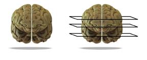
The next generation of ultrasound technology will be true volumetric images with greater diagnostic advantages. As work toward such advances continues, true volumetric image enhancement requires simultaneous (anisotropic) analysis of all available data. Figure 1, below, presents a simplistic image to be analyzed. Anisotropic analysis considers the entire brain’s volume and would include all data in Figure 1a. Isotropic analysis, however, analyzes selected planar views, such as the sections identified in Figure 1b. This type of analysis considers data from two-dimensional slices and stacks of them (multiple planar views), instead of the entire picture. The former uses three-dimensional (3D) analysis to examine a 3D image, capturing all areas within the volume.
Figure 2 presents an example in which volumetric image enhancement has been performed on the original 3D ultrasound image of the fetal brain. In the volumetrically enhanced image (on the right), structures have a clarity not visible in the original image (on the left). Several clinical evaluations have concluded that volumetric image enhancement in ultrasound adds diagnostic value, especially in cases with deep-lying organs and structures such as cysts and tumors. With 3D images, doctors can use and review the data as a volume, rather than individual slices of the volume.
Visualization of the heart in 3D is already possible and provides the physician with a tool to review its anatomical structure, identifying congenital heart anomalies or other changes. However, depicting the beating heart and performing real-time estimations to reveal information related to wall motion or other clinical information requires tremendous computational power, such as that available in multicore CPUs and other HPC platforms.
New diagnostic tools based on mathematical algorithms requiring computational power are the way of the future. The result will be fast data accessibility, which will give physicians new ways to review and explore multidimensional data, thereby improving the diagnostic outcome.
About the author: Swante Welander drives ContextVision’s new business efforts, setting the company’s strategic direction. Additionally, he monitors and analyzes the medical imaging market to allow ContextVision to meet the market’s future demands. ContextVision’s embedded imaging software enhances medical images across MRI, X-ray, ultrasound, CT, and IR.


 April 18, 2025