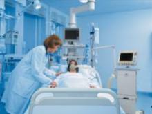
Mary Washington Hospital (MWH), part of MediCorp Health Systems in Fredericksburg, VA, has been the area’s premier healthcare provider for more than 100 years. Through the Cancer Center of Virginia, MWH offers the most advanced radiation therapy available. In 2000, they were the first community hospital in the state to introduce intensity modulated radiotherapy (IMRT), targeting tumors with greater precision and stronger doses, while offering better protection to the healthy tissue surrounding tumors.


Talk to the purveyors and supporters of digital radiography (DR) and you’ll hear that it’s the wave of the future and ultimately where the technology is headed – arguably replacing its earlier generation sibling, computed radiography (CR). However, raise that prospect with the CR crowd and you’ll likely hear something dramatically different.


It is no surprise that during the past several years, the number of magnetic resonance imaging (MRI) and computed tomography (CT) scans performed in the U.S. has dramatically escalated. An aging population and technological advances are driving these testing modalities and also forming the basis for extended offerings. In 2004 alone, the U.S. market for diagnostic imaging systems reached about $7 billion. It is expected to exceed $8 billion by 2009, according to findings by Millennium Research Group.


What are some of the noteworthy growth areas for computed tomography (CT) applications today and why are they so popular? The greatest CT growth areas are currently in cardiovascular imaging and functional studies. The ongoing evolution of cardiac CT angiography has increased the ability to visualize noncalcified lesions.


Few medical devices are credited with preventing death and are used so rarely as automated external defibrillators (AEDs), the small, portable, electronic gizmos that can restore the heart’s natural rhythm following sudden cardiac arrest.


Not too long ago, the capability of precisely directing and guiding a potent beam of radiation to eradicate a cancerous tumor without harming the healthy tissue around it was a pipe dream. Fodder for the imagination and science fiction.


One of the most prominent figures in nuclear medicine in the last half-century is Dr. Henry Wagner of Johns Hopkins University in Baltimore, MD. Dr. Wagner has conducted pioneering research in imaging the perfusion and ventilation of the lungs, kidney and spleen scanning, myocardial perfusion scanning with potassium-43, gated blood pool imaging and imaging brain receptors with PET.


Digital imaging is now ubiquitous in medicine. Contemporary imaging modalities are generating an ever-increasing amount of data, and a growing number of healthcare professionals require access to this data. However, this exponential increase in the generation and usage of digital imaging poses new data management problems for the administrators of picture archiving and communication systems (PACS). In response, emerging storage grid technologies offer a practical means to addressing many of these problems.
Explosion of Imaging Data


A 30-year-old woman arrives in the emergency room with “abdominal cramping and vaginal bleeding. A physical examination reveals a palpable left adnexal mass. She has a positive pregnancy test with beta-HCG of 14,000 IU/L. The emergency department (ED) physician considers magnetic resonance imaging (MRI) of the pelvis, yet it is expensive and not the initial study of choice.


Anyone who remembers – or for that matter, still uses – single-slice CT scanners can appreciate the dramatic advancements made by leading CT system manufacturers in recent years. The latest CT, Multidetector CT (MDCT), and MRI systems are faster and feature higher resolution than ever before.


Subscribe Now