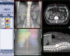
OBI integrated with Varian's VARiS Vision image and information management system and treatment planning software enables clinicians to pinpoint tumor location.
The prevalence of radiographic imaging within the oncologic department, or more specifically, within the radiation treatment room, is the end result of advancements in cancer treatment and to some degree, new knowledge of how cancer reacts to treatment.
Fundamental to any cancer treatment plan is identifying the exact location of the tumor. In years past, this accuracy, while important, was perceived as a benefit of imaging, not a necessary component. Today, with the prevalence of IMRT and IGRT, diagnostic imaging has taken its rightful place as a necessary partner during the radiation therapy treatment process.
Using imaging to gauge accuracy during radiation therapy is not a new concept. For years, linear accelerator manufacturers have addressed this issue by providing portal imagers. Yet, even advancements in Electronic Portal Imaging Devices (EPID), which provided a quantum leap in image quality and spatial resolution, could not by itself be the catalyst for further expanding the use of radiation therapy to treat tumors/cancer once labeled as “untreatable.”
In this vein, the industry recognized that increasing the accuracy of treatments was dependent upon the ability to adapt the treatment plan prior to and during the treatment process as well as recognizing the role of organ motion and tumor changes as treatment progresses. Solutions that could “personalize” cancer treatment began to emerge as the era of adaptive radiotherapy begins.
Adaptive Radiotherapy with Linear Accelerators
For adaptive radiotherapy to enable clinicians to adapt and change radiation therapy treatment to the size, shape and location of a tumor, clinicians rely upon fully-integrated imaging devices, information management systems and radiation delivery technology. Margins can be reduced to treat tumors with a previously unattained high degree of precision and accuracy, while minimizing the adverse effect of damaging surrounding healthy tissue.
Linear accelerators continue to form the foundation for adaptive radiotherapy. Elekta’s Synergy boasts an integrated 3-D volume imaging functionality that provides “CT-like” image quality. The combination of X-ray Volume Imaging (XVI) technology and reconstruction techniques are the basis for Elekta’s VolumeView volumetric 3-D data sets, which is the foundation of Elekta’s 4-D adaptive image-guided radiation therapy concept. Immediately following acquisition, the VolumeView image is available enabling the clinician to correct for organ motion or tumor size and shape changes, particularly shrinkage, that result from irradiation of the site.
Varian Medical Systems On-Board Imager (OBI) accessory for the company’s Clinac and Trilogy linacs provides 3-D cone beam CT images that show anatomic changes while the patient is on the treatment table. By integrating OBI with Varian’s VARiS Vision image and information management system and Eclipse treatment planning software, clinicians can pinpoint tumor location and minimize radiation exposure to surrounding healthy tissue.
The company’s Dynamic Adaptive Radiotherapy (DART) Initiative takes IGRT a step further with a similar integration scheme based on the company’s Inspiration platform. Beyond OBI, VARiS Vision and Eclipse, another key factor is Varian’s RPM respiratory gating system, which enables the imaging and treatment of the target site in conjunction with respiratory motion.
Dow Wilson, president of Varian’s Oncology Systems business calls DART “the next stage in personalized cancer care,” due to its ability to help clinicians adapt a patient’s treatment plan based on information from this integrated technology scheme, which accurately depicts the exact size, shape and location of the tumor just prior to dose delivery.
At Siemens Medical Solutions, ONCOR Impression is designed to integrate with Coherence Oncology Workspaces, a suite of applications that optimize workflow. Currently, Coherence Physicist is a works-in-progress within the U.S., pending FDA 510k clearance. Taking a different approach with a focus on megavoltage (versus kilovoltage with Varian’s OBI and Elekta’s Synergy) cone beam, the MVCB works-in-progress solution will produce CT-based, 3-D images for automatic, online targeting of the treatment area.
According to Ajit Singh, president, Oncology Care Systems Group, Siemens Medical Solutions, the company’s vision for adaptive radiation therapy is to “close the loop” of information at the time of treatment. This is accomplished by providing feedback to the clinicians, as images and information, during several critical points throughout the treatment process.
Adding Robotics to the Linac
When Accuray introduced the CyberKnife Robotic Radiosurgery System, the belief was that a linear accelerator with advanced robotics could provide radiosurgery precision to treat a greater array of tumors throughout the body. The concept, based on the Neurotron 1000 system still in use today at Stanford University, treats intracranial and extracranial tumors with sub-millimeter accuracy, within a one millimeter margin of error according to the manufacturer. With image guidance technology and computer controlled robotics, the CyberKnife System continuously tracks, detects and corrects for tumor and patient movement throughout the treatment. The system’s Synchrony respiratory tracking system correlates motion by graphing the tumor’s position by imaging fiducial markers.
According to Iris Gibbs, M.D., co-director of CyberKnife Radiosurgery Program and assistant professor, Radiation Oncology, Stanford University, “The CyberKnife linac is attached to a commercial robot to provide greater movement and achieve angles that fixed gantries cannot attain.”
Image is Everything
The importance of linacs notwithstanding, diagnostic images is driving precise tumor localization.
Advances in tumor localization have reached a new level at University of Washington, Seattle, where 4-D whole-body respiratory gated PET imaging is now routine. Using the GE Discovery STE PET/CT, Kinahan demonstrated how the scanner allows for simultaneous acquisition of whole-body respiratory gated PET and a list mode coincidence file. As a result, “4-D PET has been acquired as part of the clinical routine and is currently conducted on all whole-body PET/CT exams,” indicated Paul Kinahan, associate professor of Bioengineering, University of Washington. In addition, respiratory motion of the lung, liver and heart can be captured and assessed across the entire patient FOV. “We believe 4-D will be used routinely by many clinicians,” added Molecular Imaging Leader Jean-Luc Vanderheyden, GE Healthcare.
GE Healthcare’s Advantage SIM MD provides 4-D multiplanar motion visualization using images from multiple modalities, including CT, PET and MRI. “The software captures the full range of motion of critical internal structures and lesions during respiration, providing a more accurate description of the target and its trajectory,” said Gene Saragnese, vice president and general manager, Global Functional and CT, GE Healthcare.
By providing a real-time view of anatomy in motion, clinicians can select an appropriate respiratory cycle in the treatment plan. This can minimize exposure of healthy tissue to radiation, while helping oncologists optimize the use IGRT and IMRT treatment methods.
Integration is the Key
Regardless of hardware or software, the unifying concept found in each vendor’s technology is the integration of images and information at the point of care. The ability to adapt opens the door to more than personalized radiation therapy; it also pushes the envelope for successfully and safely treating tumors in numerous areas of the body once deemed untreatable with external beam radiation, including the spine, lung, liver and prostate.
As molecular imaging continues to uncover tumor response to treatment, biological information will also be integrated into this schematic. Clinicians will then understand the unique qualities of the disease within each patient and tailor a treatment plan to their unique molecular and biological composition.