
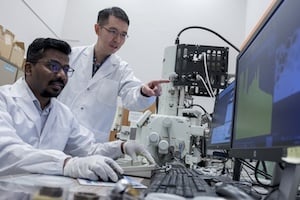
Associate professor Wong Liang Jie (left) and Research Fellow Dr. Nikhil Pramanik from NTU Singapore prepare a sample to be analyzed using a field emission scanning electron microscope for their research team’s X-ray experiments. The team found a new way to produce water-window X-rays, which could make bioimaging X-ray machines smaller and more flexible to use. (All photos: NTU Singapore)
Water-window X-rays allow researchers to visualize biological cells at high contrast without staining agents or other potentially damaging preparations. Multiple challenges, however, have prevented widespread adoption.
Previously, the only way to produce water-window X-rays of varying energies was with synchrotrons, expensive facilities that are the size of a large stadium. Most smaller tabletop machines could only produce radiation in fixed energies, and some required high-intensity lasers that need ongoing maintenance.
Now, researchers at Nanyang Technological University, Singapore (NTU Singapore) have developed a method to produce water-window X-rays of varying energies using a single, table-sized setup, a breakthrough that could expand the use of the technology worldwide.1
“Compared to a synchrotron, our table-sized setup has a potentially smaller carbon footprint, smaller physical footprint, and is more accessible and less expensive for lab researchers to use,” says associate professor Wong Liang Jie from NTU Singapore’s School of Electrical and Electronic Engineering, who helped develop the technology.
What Makes Water-Window X-rays Different?
X-rays for medical diagnostics pass through both water and carbon-rich biomolecules in cells, making it difficult to distinguish between them. In contrast, water-window X-rays operate at wavelengths between 2.3 and 4.4 nanometers (nm), allowing researchers to see living cells and their structures clearly. “This is important in certain studies, such as capturing visuals of viruses attacking cells in real time,” Wong says.
Some researchers visualize cell structures at high resolution with transmission electron microscopy (TEM), which uses electrons to create images. “But the preparation needed, such as slicing samples into ultrathin sections, often means only dead samples can be used,” Wong explains. “With water-window X-rays, living cells can be imaged.”
Producing Tabletop Water-Window X-rays
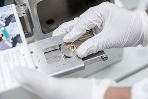
To generate water-window X-rays in the lab at NTU, researchers fired low-energy electrons at ultrathin flakes of graphite 10 nm to 170 nm thick. This technique energized the graphite atoms, causing them to emit X-rays.
“The atoms of the graphite flakes were arranged in a very orderly way, with a repeating pattern, like a crystal,” Wong explains. “This structure makes them behave like an array of nanoantennas that work together to send out only X-rays of a specific wavelength in a specific direction, while filtering out radiation of other wavelengths and directions.”
Wong and his team adjusted their setup so water-window X-rays are the dominant radiation produced. “In our setup, the electrons fired at the graphite flakes need to be sped up first,” Wong says. “But since our technique requires electrons of low energy, we don’t need to accelerate the electrons too much, so we required a setup about the size of a table to do so.”
The tunability of the setup, Wong says, helps exploit the fact that different elements, such as carbon and oxygen, absorb water-window X-rays of different energies to different extents. This allows for greater contrast, helping researchers distinguish different biomolecules and cell structures more easily.
Potential Implications for Researchers, Hospitals
After performing proof-of-concept experiments with modified electron microscopes, researchers at NTU Singapore are now assembling a prototype for a full-fledged imaging device to showcase commercially competitive results.
Wong believes that basic research will benefit first from a tabletop water-window X-ray setup. “But pathology, drug development, and even X-ray imaging for non-bio-related applications like solar cells, batteries, and semiconductors could be impacted the most later because they are technologies we use in everyday life.”
Could water-window X-rays eventually be used in a hospital setting? “They would be invaluable for performing high-resolution, 3D biopsies and studying how human cells interact with pathogens and viruses,” Wong says. “This is especially relevant today, where climate change and global warming have been triggering new waves of disease outbreaks.”
Getting to that point, however, could take years, Wong surmises. “Extensive testing, engineering, and tweaking are still needed to develop and optimize a commercially viable product,” he says.
Reference
-
Pramanik, Nikhil & Huang, Sunchao & Duan, Ruihuan & Zhai, Qingwei & Go, Michael & Boothroyd, Chris & Liu, Zheng & Wong, Jie. (2024). Fundamental scaling laws of water-window X-rays from free-electron-driven van der Waals structures. Nature Photonics. 18. 1203-1211. 10.1038/s41566-024-01547-3.

 June 19, 2026


