June 24, 2023 — A new ultra-high resolution (UHR) brain PET scanner may have the ability to characterize previously indistinguishable brain regions that are known to be involved in Alzheimer’s disease, depressive disorders, visual attention disorders, tinnitus, and other conditions. With its unprecedented resolution, the UHR brain scanner opens the door to more accurate studies of the brainstem. This research was presented at the Society of Nuclear Medicine and Molecular Imaging 2023 Annual Meeting.
“Up until now, PET has been useful for the study of neurological phenomena and for diagnostic purposes, but its potential has been somewhat limited by the poor spatial resolution of current PET systems,” said Vincent Doyon, a master’s student in Radiation Sciences and Biomedical Imaging at the 8 in Québec, Canada. “Amid technological advances in PET instrumentation and a global thrust to extend brain PET imaging capabilities, our team at the Sherbrooke Molecular Imaging Center has designed and built the UHR dedicated brain PET system.”
Unlike other PET scanners, the UHR scanner features truly pixelated detectors and reaches a 1.25 mm isotropic spatial resolution, a two-fold improvement over the High Resolution Research Tomograph (HRRT) scanner, which has been considered the state-of-the-art reference for brain PET imaging for the past 20 years. “This makes it possible to visualize for the first time the radiotracer uptake in the range of a few tens of microliters in the human brain,” points out Roger Lecomte, PhD, professor of Nuclear Medicine and Radiobiology at University of Sherbrooke.
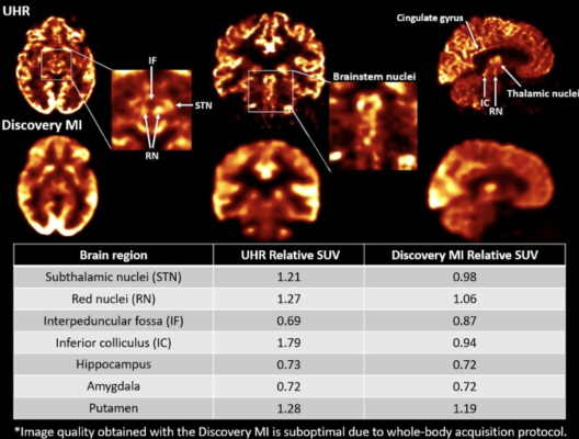
To demonstrate the ability of the UHR scanner to delineate small cerebral structures and accurately quantify the in vivo tracer concentration, researchers compared the UHR scanner to a whole-body PET scanner. Three patients who were prescribed an 18F-FDG PET scan underwent their clinical examination on the whole-body PET scanner followed by a brain scan on the UHR scanner. Images from the two scanners were compared, and region identification was performed. Standardized uptake values relative to the cerebellum were also calculated.
Several regions of the brain were easily identified visually in UHR images (particularly in the brainstem) but could not be visualized by the whole-body PET scanner. Inferior and superior colliculi as well as the subthalamic nuclei and red nuclei were clearly delineated in the UHR images. In addition, the thalamus—which is commonly seen as a whole in standard PET images—could be visually segmented into smaller nuclei with the UHR scanner. Hypermetabolic regions of the cortex were also noticeable in the UHR images but were hardly perceivable with the whole-body PET scanner.
“The UHR scanner is a quantum leap for PET image resolution,” Doyon stated. “Proper visualization of brainstem nuclei will provide the ability to detect early changes associated with many diseases and offer a potential avenue for early diagnosis. This will impact both research and clinical settings.”
The first UHR prototype is fully operational and is used for research applications at the Sherbrooke Molecular Imaging Center. Within the next several months, additional UHR units will be deployed to brain research centers across North America while progressing towards obtaining the necessary regulatory approvals for clinical imaging. The performance of a second UHR model with new detectors improving the overall resolution across the field-of-view will also be tested soon.
For more information: www.snmmi.org
Find more SNMMI23 conference coverage

 October 08, 2026