Image courtesy of the University of Minnesota Medical School
June 2, 2023 — The University of Minnesota and Te Herenga Waka—Victoria University of Wellington, NZ, have received National Institutes of Health (NIH) grants, totaling over $12 million, to work on an international, multi-institutional project to develop a radically new magnetic resonance imaging (MRI) scanner that is compact and transportable.
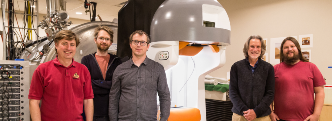
“Over the past eight years, with funding from the NIH BRAIN Initiative and other sources, we have created MRI technology that allowed us to develop a prototype brain MRI scanner that promises to expand existing MRI capabilities and accessibility,” said study principal investigator Michael Garwood, PhD, a professor in the U of M Medical School.
The MRI scanner is a technology that allows doctors and scientists to see detailed pictures of the human brain. It is useful in understanding how the brain works and diagnosing medical conditions. Today, it is essential in the fields of neuroscience and clinical medicine. Yet, despite its tremendous importance, most of the world’s population cannot access MRI.
MRI machines are expensive and require specialized facilities and trained professionals to operate and maintain them. In addition, their large size and weight make it difficult to take them to remote or resource-limited areas. As a result, MRI is primarily available to the middle and upper classes in wealthy countries.
“The design of our new MRI head-only magnet, developed by the team at Te Herenga Waka in New Zealand, will allow studies of the human brain without a highly confining MRI magnet bore,” Dr. Garwood said. “Due to its small size and its light weight, this MRI scanner will be transportable for imaging brain function and structure almost anywhere, for almost anyone”
“Future portable MRI scanners like ours may empower communities in remote, resource-limited settings to address health inequities, perform research leading to improved understanding of brain development and degeneration in diverse populations, and improve access to quality clinical care,” said Dr. Garwood.
In the Center for Magnetic Resonance Research at the U of M, the research team plans to use MRI techniques to create different types of images that are needed for various research purposes. After that, they will collaborate with communities that historically have had limited access to medical resources, such as Native American tribal communities, to conduct a pilot study of this technology in the field.
Ben Parkinson, a senior engineer at Te Herenga Waka, said collaboration between countries and universities has been seamless. “Developing a device that needs to work together both mechanically and electromagnetically would be a complicated enough endeavor within your own institution, let alone internationally. But, the team has been very successful, and I am excited to continue our partnership.”
Funding for this culmination of grants is provided by NIH BRAIN Initiative Grant Numbers: R24 MH105998-01 and U01 EB025153-01. The first grant provided funds to plan and investigate the feasibility of this groundbreaking MRI system. The second grant is currently in a no-cost extension, provided the funds to build it.
For more information: www.med.umn.edu

 July 20, 2026


