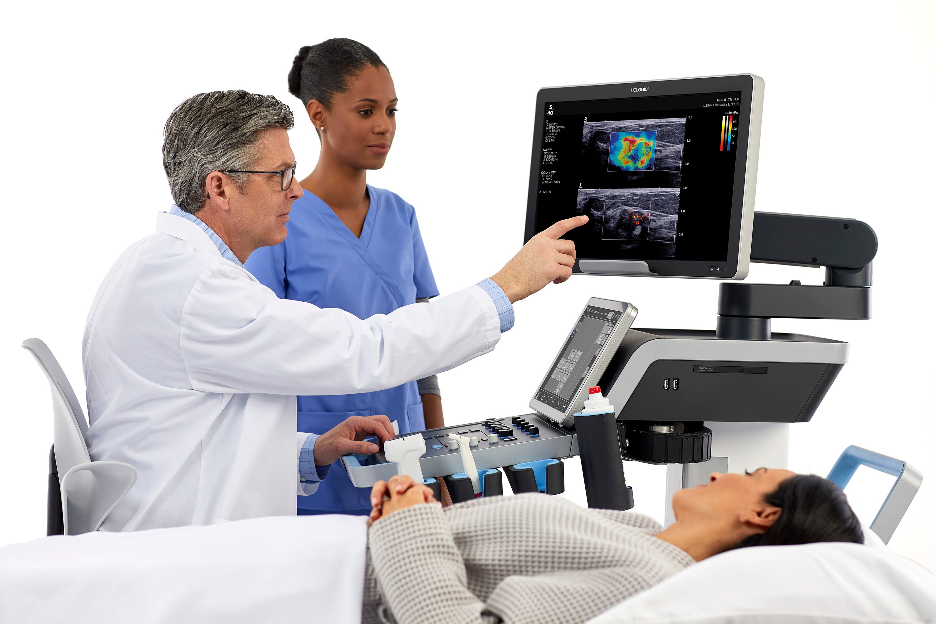
When performing a biopsy procedure, radiologists can feel more confident in their sampling with the assistance of shear wave elastography. Image courtesy of Hologic
Ultrasound has become one of the most important diagnostic tools in a radiologist’s toolkit. In the early days of ultrasound imaging, it was designed to provide an anatomical and morphological view of the patient. However, as the technology has continued to improve, today ultrasound can provide more detail and information, including visualizing tissue stiffness, microvascularization, the level of fat content in the tissue and inflammation. These biomarkers are now critical to a radiologist’s imaging analysis.
Ultrasound is an important modality in breast cancer diagnosis as it is typically the next step following the discovery of an abnormality through a mammogram. Particularly for women with dense breast tissue, ultrasound can provide an essential view of lesions to escalate or downgrade their risk of cancer by differentiating healthy tissue from a suspicious mass.

The color-coded mapping of breast lesion stiffness is a complementary tool for breast ultrasound that provides additional information that can help radiologists feel more confident in their diagnosis. Image courtesy of Hologic.
When innovative imaging modes like shear wave elastography are combined with ultrasound, it may aid in the diagnostic work up of breast lesions. Shear wave elastography builds on the latest imaging technology to offer complementary diagnostic information. While it has made many strides since it was first introduced to ultrasound imaging, many radiologists have not yet been exposed to the benefits of this modernized technology and thus not adopted it into their everyday practice.
However, with more than 200 peer-reviewed publications affirming the positive impact of shear wave elastography solutions for the breast, this is one technology that radiologists should consider as part of their ultrasound workflow. It can be a key element in diagnosis by optimizing lesion characterization and reducing unnecessary biopsies.1 In addition, shear wave elastography can aid radiologists in effectively and efficiently identifying cancerous lesions, enhancing ultrasound guided biopsies, and treatment planning and monitoring through real-time data, acquired noninvasively and easily, that aligns with ultrasound procedures.
Lesion Characterization
Shear wave elastography is a real time technique capable of noninvasively measuring tissue stiffness to make ultrasound exams more efficient for the radiologist to best manage their patients’ clinical pathway. This technology can help radiologists accurately view and diagnose lesions by analyzing its stiffness in real time on top of their conventional ultrasound exam. The color-coded mapping of breast lesion stiffness is a complementary tool for breast ultrasound that provides additional information that can help radiologists feel more confident in their diagnosis and has the potential to reduce benign breast biopsies and false negatives.
When shear wave elastography is added to B-mode during the ultrasound exam, radiologists have reported being able to add a critical biomarker to their routine to downgrade more lesions from BI-RADS 4a to BI-RADS 3 to avoid unnecessary biopsies. Additionally, radiologists can measure the histopathologic severity of breast masses through the technology in order to monitor the lesion in follow-up exams. By including shear wave elastography in their clinical routine, radiologists can optimize the accuracy and efficiency of breast ultrasound to help patients find comfort in the accuracy of their diagnosis and the path forward.
For radiologists, this imaging technology provides efficient patient management as well, which helps promote workflow. Shear wave elastography offers improved specificity of conventional breast ultrasound exams that maintains sensitivity to better identify if a mass is malignant or benign. By utilizing shear wave elastography, radiologists can have more clarity on lesion characteristics, and can even note a more accurate lesion size measurement.2 These fast results enable radiologists to not only accurately diagnose, but also potentially improve their time management for productivity and react more quickly according to patient state.
Ultrasound Biopsy Guidance
When a patient needs to undergo a biopsy procedure, radiologists can feel more confident in their sampling with the assistance of shear wave elastography. The added value of real-time shear wave elastography to the medical imaging landscape is recognized by major scientific societies in ultrasound3 since the technology provides additional imaging information about its stiffness. These details can potentially improve the procedure’s success as shear wave elastography has been shown to help radiologists target lesions more efficiently during guided biopsy4 due to its ability to help radiologists visualize, analyze and quantify tissue stiffness in real time. Through this technology, the stiffest area within the mass is color-coded on the mapping system helping to target samples to acquire more accurate specimens.
In some instances, shear wave elastography has been proven to help radiologists detect more cancer cells during the biopsy, which enables them to make more informed decisions for patient care and treatment.
A patient’s experience with biopsy can be positively impacted by the tools and equipment used to gather a sample. By utilizing shear wave elastography, radiologists can provide a better experience while also adding value to their medical imaging landscape.
Treatment Planning and Monitoring
Through the prognostic information provided by the shear wave elastography real time mapping, radiologists can determine next steps in the patient’s care, such as monitoring the pathological response during and after neoadjuvant chemotherapy before surgery.
Shear wave elastography’s real time mapping of breast lesion stiffness provides additional diagnostic information to improve the management of patients through the breast health journey. Throughout this process, the technology enables radiologists to meet expectations for patients confidently and deliver informed pathology plans for treatment.
This imaging technology provides the vital information radiologists need to determine if treatment has been successful. By measuring lesion stiffness, shear wave elastography provides a unique view of the breast that can provide critical information as patients undergo treatment. Should surgery be needed, shear wave elastography can also be used to ensure cancerous lesions are completely removed from the breast tissue, and to monitor patients in the months and years that follow.
Liver Application
Just as ultrasound can be used for a variety of needs throughout the body, so can shear wave elastography. By driving innovation within the ultrasound landscape, shear wave elastography can add clinical value for radiologists and more efficiently address the needs of patients.

An investment in shear wave elastography is not just in breast health, as it can be utilized in other clinical applications, such as liver disease.
An investment in shear wave elastography is not just in breast health, as it can be utilized in other clinical applications. Shear wave elastography provides innovative solutions to diagnose and manage patients suffering from chronic liver diseases like severe alcoholic steatohepatitis, nonalcoholic steatohepatitis and nonalcoholic fatty liver disease. This imaging technology provides radiologists with additional detailed information to make confident, accurate diagnoses. Through its easy-to-use, color-coded mapping system, radiologists can quickly assess the stiffness of the liver, and when overlapped with other ultrasound data, can help them determine the necessary next steps for a patient.
It is critical to diagnose chronic liver diseases early because most patients do not suffer from symptoms until they reach an advanced form, which can lead to more complications, portal hypertension, esophageal varices, cirrhosis and liver cancer. Through ultrasound imaging with shear wave elastography, radiologists can identify new ultrasound biomarkers to diagnose their patients earlier and manage treatment efficiently.
This innovation is driving to one goal, no matter the application: early detection. Across the medical field, early detection is key to improving patient access to the best treatments. Shear wave elastography is a vital tool in the radiologist’s toolkit that advances this mission. By positively impacting patient management and supporting diagnostic decision making, this imaging technology enables radiologists to deliver the best care possible. When radiologists are more confident in their diagnosis, patients can feel less anxiety about the next steps of their care.

Jennifer Meade is the President of Hologic’s Breast and Skeletal Health Division and has been part of the company since 2006. In her role, she is committed to continuing to drive innovation across the breast care continuum and investing in the company’s people to ultimately drive improved outcomes for patients around the globe.
References:
1. Berg WA et Radiology 2012 Feb;262(2):435-49.
2. Mullen R et Clin Radiol 2014 Dec;69(12):1259-63.
3. ACR BI RADS: Ultrasound (2015), KSUM Guidelines for breast elastography (2014), EFSUMB (2013) and WFUMB Guidelines
4. Plecha DM et Radiology 2014; 272(3):657-64.
Related Shear Wave Elastography Content:
Preoperative Shear Wave Elastography Helps Predict Rotator Cuff Repair
Shear-Wave Elastography for Interventional Monitoring of Pediatric Budd-Chiari Syndrome
New Cancer Scan Could Guide Brain Surgery
Studies Confirm Clinical Value of ShearWave Elastography for Liver Fibrosis Evaluation

 July 22, 2026


