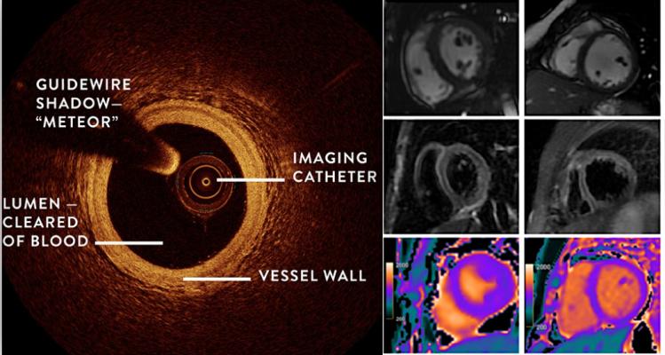
The Women’s Heart Attack Research Program (HARP) study shows combining OCT and cardiac MRI can help detect the underlying cause of heart attacks in women who did not have blocked arteries.
April 30, 2021 — In almost 10 percent of myocardial infarctions, no obvious cause in the coronary artery can be found. Some of the patients are diagnosed with broken-heart syndrome, while others are left without a diagnosis. A new study from Karolinska Institutet in Sweden suggests that early magnetic resonance (MRI) imaging of the heart can greatly increase the rate of diagnosis. The study has been published in the journal JACC Cardiovascular Imaging.
Myocardial infarction is one of the most common diseases in the West, and is usually caused by a blood clot that blocks the coronary artery on the heart's surface. However, in up to ten per cent of all myocardial infarctions, no obvious cause in the coronary artery is found. Such patients are given the working diagnosis MINOCA (myocardial infarction with non-obstructive coronary arteries), which can subsequently lead to one of several diagnoses.
The majority of the patients are women, many of whom are diagnosed with takotsubo cardiomyopathy (broken-heart syndrome). This is a condition characterised by reduced heart function that is probably related to stress and that presents the same symptoms as a regular heart attack.
"Around 80 to 90 per cent of broken-heart sufferers are women, and the disease is associated with mental stress," said principal investigator Per Tornvall, senior physician and professor at the Department of Clinical Science and Education, Södersjukhuset (Stockholm South General Hospital), Karolinska Institutet. "There also seems to be a link to hypersensitivity towards stress caused by low oestrogen levels. Unfortunately, research on the investigation and treatment of myocardial infarction is often done on men, while female heart disease is less studied."
Cardiovascular magnetic resonance (CMR) is often done when examining patients with MINOCA. CMR approximately ten days after onset can result in a diagnosis in under half the patients, normally takotsubo or myocarditis (inflammation of the heart muscle), according to an earlier study of 150 patients from Karolinska Institutet. The same researchers have now tested a new, more sensitive CMR technique two to four days after onset on a comparable group of 148 patients with a median age of 58. They found that a full 77 percent of the patients could be given a diagnosis (35 per cent of takotsubo and 17 per cent of myocardial inflammation, compared with 19 and 7 percent, respectively, in the first study).
"We don't know how much effect the improved CMR technique has, but the results suggest that with early examination more patients can get a correct diagnosis and therefore the right treatment," said Professor Tornvall. "The next step is for us to develop the CMR examination with pharmacological stress of the heart. This will enable us to study the smallest of the blood vessels and hopefully find a cause for the 23 per cent who received no diagnosis."
The study was conducted in close collaboration with the heart-MR department, clinical physiology, at Karolinska University Hospital and was financed by the Swedish Research Council, Region Stockholm and the Swedish Heart and Lung Foundation. Co-authors Peder Sörensson, Magnus Lundin and Martin Ugander are affiliated with Karolinska University Hospital, which has a research agreement with Siemens Healthineers regarding CMR. Jonas Spaak has received personal fees for the past three years from AstraZeneca, Vifor Pharma and Novo Nordisk, all unrelated to the present study.
For more information: https://ki.se/en
 October 23, 2026