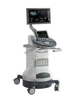
July 31, 2019 — SuperSonic Imagine announced the publication of the results of its prospective multicentric clinical study conducted in China to validate the performance of ShearWave Elastography (SWE) in evaluating liver fibrosis severity in patients with chronic hepatitis B (HBV). SWE enables tissue stiffness to be instantly visualised and measured in kPa. The results of the study were published in two articles that appeared in the peer-reviewed journals, Radiology1 and Gut2.
This study collected data from 12 Chinese level 3 hospitals from January 2015 to January 2016. All of the 402 patients included received the same prospective protocol, including a liver biopsy, an SWE examination and a blood test. Some were also assessed with VTCE (transient elastography). All images, measurements and histopathological analyses were subjected to centralized quality control, thus ensuring data reliability and consistency.
The analysis published in Radiology in July 2018 followed the recent recommendations of the European Association for the Study of the Liver (EASL) pertaining to the care of patients infected with the hepatitis B virus. The patients were divided into two groups: a group with chronic inactive disease, who receive regular monitoring; and a group with active chronic hepatitis who require treatment with antiviral drugs.
Results confirm that SWE is superior to other non-invasive tests to assess fibrosis severity, particularly in cases of cirrhosis. In practice, for patients with a chronic inactive hepatitis B infection, a stiffness above 11 kPa could lead to the diagnosis of cirrhosis and thus identifying patients requiring treatment. In contrast, a stiffness below 8.5 kPa could exclude the diagnosis of cirrhosis thus identifying patients requiring monitoring. Although a liver biopsy would still be recommended for a stiffness between 8.5 and 11 kPa, the study demonstrates the SWE examination could have avoided the need for biopsy in 81.2 percent of patients (125 out of 154).
“This prospective multicentric study confirmed that two-dimensional SWE is superior to other non- invasive methods in the diagnosis of liver fibrosis and cirrhosis. It also demonstrated that 81.2 percent of patients with chronic HBV infection could avoid undergoing a biopsy using SWE.” stated Prof. Ping Liang, M.D., principal investigator of the study, from the People’s Liberation Army Hospital in Beijing.
The article published in British gastroenterology journal Gut reports the results of the use of radiomic and neural network techniques to extract quantifiable features from SWE images (DLRE) to predict fibrosis severity.
The results of this analysis are very promising, given that the diagnostic performance of DLRE was found to be equivalent to the gold-standard biopsy for determining stages of fibrosis. The DLRE technique could therefore represent an initial step towards standardizing practices between radiologists and clinicians, avoiding operator measurements, and encouraging the adoption of SWE and DLRE by non-radiological users.
For more information: www.supersonicimagine.com
References
1. Chauhan A. The Emerging Role of Two-dimensional US Shear-Wave Elastography in Chronic Liver Disease. Radiology, published online July 24, 2018. https://pubs.rsna.org/doi/10.1148/radiol.2018181281
2. Wang K., Lu X., Zhou H., et al. Deep learning Radiomics of shear wave elastography significantly improved diagnostic performance for assessing liver fibrosis in chronic hepatitis B: a prospective multicentre study. Gut, May 5, 2018. https://gut.bmj.com/content/early/2018/05/04/gutjnl-2018-316204

 May 27, 2026


 Clearance")


