October 18, 2017 — A machine learning tool can help identify which high-risk breast lesions are likely to become cancerous, according to a new study appearing online in the journal Radiology. Researchers said the technology has the potential to reduce unnecessary surgeries.
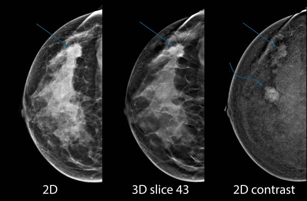
High-risk breast lesions are biopsy-diagnosed lesions that carry an increased risk of developing into cancer. Because of that risk, surgical removal is often the preferred treatment option. However, many high-risk lesions do not pose an immediate threat to the patient's life and can be safely monitored with follow-up breast imaging, sparing patients the costs and complications associated with surgery.
"There are different types of high-risk lesions," said study author and radiologist Manisha Bahl, M.D., M.P.H., from Massachusetts General Hospital (MGH) and Harvard Medical School, both in Boston. "Most institutions recommend surgical excision for high-risk lesions such as atypical ductal hyperplasia, for which the risk of upgrade to cancer is about 20 percent. For other types of high-risk lesions, the risk of upgrade varies quite a bit in the literature, and patient management, including the decision about whether to remove or survey the lesion, varies across practices."
Bahl and colleagues at MGH studied the use of a machine learning tool to identify high-risk lesions that are at low risk for upgrade to cancer. The study resulted from a close collaboration between researchers at the Massachusetts Institute of Technology's (MIT) Computer Science and Artificial Intelligence Laboratory in Cambridge, Mass., and breast imaging experts at MGH.
"Because diagnostic tools are inexact, there is an understandable tendency for doctors to over-screen for breast cancer," said coauthor Regina Barzilay, Ph.D., the Delta Electronics Professor of Electrical Engineering and Computer Science at MIT. "When there's this much uncertainty in data, machine learning is exactly the tool that we need to improve detection and prevent overtreatment."
Machine learning is a type of artificial intelligence in which a model automatically learns and improves based on previous experiences. The model developed by researchers analyzed traditional risk factors such as patient age and lesion histology, along with several unique features, including words that appear in the text from the biopsy pathology report. The researchers trained the model on a group of patients with biopsy-proven high-risk lesions who had surgery or at least two-year imaging follow-up. Of the 1,006 high-risk lesions identified, 115, or 11 percent, were upgraded to cancer.
After training the machine learning model on two-thirds of the high-risk lesions, the researchers tested it on the remaining 335 lesions. The model correctly predicted 37 of the 38 lesions, or 97 percent, that were upgraded to cancer. The researchers also found that use of the model would have helped avoid almost one-third of benign surgeries.
The machine-learning model identified the terms "severely" and "severely atypical" in the text of the pathology reports as associated with a greater risk of upgrade to cancer.
"Our study provides proof of concept that machine learning can not only decrease unnecessary surgery by nearly one-third in this specific patient population, but also can support more targeted, personalized approaches to patient care," said the paper's senior author, Constance Lehman, M.D., Ph.D., professor at Harvard Medical School and Director of Breast Imaging at MGH.
"Our goal is to apply the tool in clinical settings to help make more informed decisions as to which patients will be surveilled and which will go on to surgery," Bahl added. "I believe we can capitalize on machine learning to inform clinical decision making and ultimately improve patient care."
Watch the VIDEO “Examples of Artificial Intelligence in Medical Imaging Diagnostics.”
For more information: www.pubs.rsna.org/journal/radiology
 October 09, 2026