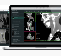
As research and development continues to reveal the benefits of medical imaging in healthcare, vendors are steadily improving imaging scanners for added efficiency and workflow. Because advanced visualization technologies are a part of the larger medical imaging discussion, these products are also evolving and growing in response to innovation within medical imaging.
Market Leaders
GE Healthcare, Siemens Healthcare, Vital Images, TeraRecon and Philips Healthcare continue to lead the advanced visualization market. In KLAS’ recent research report, “Advanced Visualization 2013: How Advanced Is It?” Philips led in overall performance with a score of 86.2. GE, TeraRecon, Vital Images and Siemens followed with scores of 84.5, 83.3, 82.5 and 79.4, respectively.1
According to the report, however, providers are still looking for more complete advanced visualization systems with added functionality in the top three critical areas — cardiac, vascular and neuro imaging. None of the top vendors are listed as the leader in functionality in all three areas — a need that is currently preventing advanced visualization software from being used as a primary reading environment. “Two-thirds of providers said they would consider using the advanced visualization software for their primary reading environment, but only if and when complete functionality is offered,” the report stated.1
Recent Software Releases
Smaller vendors have tried to address this need, introducing various applications within the past year. At the 2014 Healthcare Information and Management Systems Society (HIMSS) conference in February, Penrad released its Visualize:Vascular software, which generates 3-D images of the carotid artery lumen from slides created by conventional 2-D ultrasound.
Additionally, Aycan and Chimaera GmbH received U.S. Food and Drug Administration (FDA) 501(k) clearance for the Chimaera FusionSync software, which the company said allows for more efficient handling and diagnosis of multi-modal and follow-up medical data. The software supports 3-D computed tomography (CT), C-arm CT, magnetic resonance imaging (MRI), positron emission tomography (PET) and single-photon emission computed tomography (SPECT) data. EOS Imaging also gained CE mark this year for its hipEOS — a 3-D hip arthroplasty planning software that uses the company’s biplanar 3-D imaging.
In April, Shina Systems also announced the new commercial release of its 3Di picture archive and communications system (PACS). According to the company, the system allows clinicians and patients to view medical images anywhere.
Global Expansion
Advanced visualization vendors are also expanding to markets outside of the United States. In addition to collaborating with Imperial College London to bring High Definition Volume Rendering (HDVR) to minimally invasive robotic surgery, Fovia Medical Inc. is also collaborating with Visua Medica to develop high-quality, accessible and affordable advanced visualization products to meet Latin America’s numerous medical imaging sub-markets.
Blackford Analysis and ContextVision have also expanded their presence in international markets. At the 2014 Arab Health Conference in Dubai, Blackford Analysis introduced its software products to the Middle Eastern market. ContextVision expanded its presence in the Chinese market by adding an application specialist to its Beijing office in order to meet high demands in the region.
New Applications for Healthcare
As more advanced visualization techniques and applications are introduced, they are finding use in many different areas of medicine. Research presented at the annual meeting of the Society of Interventional Radiology (SIR) in March found that using 3-D MRI scans to precisely measure living and dying tumor tissue before and after chemoembolization — chemotherapy targeted directly at tumors — better predicts patient survival. “Our high-precision, 3-D images of tumors provide better information to patients about whether chemoembolization has started to kill their tumors so that physicians can make more well-informed treatment recommendations,” said Jean-Francois Geschwind, M.D., Johns Hopkins interventional radiologist and senior investigator on the studies.
Engineers from the University of Washington (UW) created a new advanced imaging technology that allows scientists to see within small blood vessels to prevent injuries during cosmetic procedures. “Filler-induced tissue death can be a really devastating complication for the patient and provider,” said Shu-Hong (Holly) Chang, M.D., a UW assistant professor of ophthalmology specializing in plastic and reconstructive surgery. “This noninvasive imaging technique provides far better detail than I’ve ever seen before and helped us figure out why this is happening.” The technology, called optical micro-angiography, works similarly to ultrasound and produces 3-D images of the body’s vascular network by shining a light on the tissue.
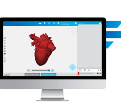
3-D imaging is also helping with personalized surgery in newborn and infant patients. 3D Systems’ Medical Modeling Virtual Surgical Planning (VSP) technology was used to aid in planning and conducting a lower jaw lengthening procedure on a young child in New York. VSP captures a digital model of the patient from an MRI or CT scan, and modeling experts work with physicians to create a surgical plan and print 3-D models and surgical guides. “VSP has not only helped make surgical procedures more precise, but offers the potential to change the scope of what is surgically possible,” said Oren Tepper, M.D., attending surgeon, Division of Plastic and Reconstructive Surgery, Montefiore Medical Center, New York. itn
Reference:
1. M. Terry. “Advanced Visualization 2013: How Advanced Is It?” KLAS, July 2013.


 April 18, 2025


