High-resolution CE-MRA carotids using Philips Sense and Centra.
When Claude Bernard inserted a mercury thermometer into the carotid artery of a horse to measure blood temperature in 1844, little did he know he was laying the foundation for present day catheter angiography. Even though his practices were rudimentary compared to today’s standards, and it was not until 1929 that basic radiographic methods were implemented, he provided a stepping-stone into the current world of catheters and in turn, vascular imaging. Catheter angiography has been the benchmark for vascular imaging because of its ability to image blood vessels invasively, technological advancements have made vascular assessment possible with a noninvasive twist.
Advancing procedures such as CT angiography (CTA), MR angiography (MRA) and vascular ultrasound offer new efficiencies in vascular imaging. Each of the modalities has its advocates, and with time each of them will continue improving, making radiologists more effective in the diagnosis of vascular diseases.
CTA Outpaces Standard Care
CTA, a noninvasive procedure that uses X-rays to image blood vessels throughout the body, is on its way to proving to be a faster and more cost-effective method to detecting coronary heart disease.
In a recent study led by Gilbert Raff, M.D., director of Cardiovascular MRI and CT Research at William Beaumont Hospital in Royal Oak, MI, CTA demonstrated that it could definitively and more rapidly exclude coronary artery disease as the cause of acute chest pain than the standard of care evaluation, including serial cardiac enzymes, ECGs and sestamibi rest-stress nuclear scanning. Among the 200 patients included in the study, length of stay in the emergency department decreased by 43.4 percent as CTA took 12.5 hours, while standard care averaged 22.1 hours.
“If I summarize the depositions we are able to achieve the high-spatial resolution while keeping it isotropic and improving the coverage, basically making it superior to all other modalities,”1 said Ergodan Cesmeli, marketing manager Cardiovascular, Molecular and CT Imaging, GE Healthcare.“ Plus CTA being mutually superior to X-ray from a radiation dose point of view, as well as providing 3-D view and providing nonvessel structure, it is continuing to be advantageous.“
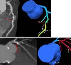
Another advantage is that anatomical detail of blood vessels can be viewed in 3-D using CTA, which can be very helpful for prevention. These preventative abilities are key when addressing steadily growing numbers of vascular disease.
There are risks, albeit minimal, associated with CTA, since the procedure uses X-rays and contrast material. “The contrast that we use is nonionic, which means that it contains no salts so the allergic risks related to it are extremely minimal,“ said Claudio M. Smuclovisky, M.D., director South Florida Medical Imaging, Boca Raton, FL. “We have done over 3,000 patients and we have never had a significant complication other than a rash that can be easily treated with a steroid or antihistamine, so the risks are extremely low.”
Although the radiation dose from the X-rays should not be considered dangerous, Dr. Smuclovisky indicates that the procedure on any given individual should be kept to a minimum and should not be done annually.
MRA Breaks into Multiphase Imaging
One benefit of MRA is that it is a noninvasive procedure, using magnetic resonance instead of radiation. Although it requires contrast material, it is gadolinium-based — much safer than others and is used in much smaller doses.
“Typically, MRA uses 20 or 30 cc’s of gadolinium injected intravenously compared to 100 or 150 cc’s done in CT or X-ray angiography,” said Stuart Clarkson, cardiovascular MR product development manager, GE Healthcare.
Like CTA, MRA has the ability to acquire 3-D images, but within the last three or four years, MRA has begun doing multiphase imaging, a method unique to MR. Multiphase imaging takes repeated images of an area as the contrast material travels through the veins. This is difficult to do with CTA because it would have to continually repeat X-rays, and there would be a risk with the radiation dose. Clarkson explained that this could be particularly useful in studies of the lower legs where blood flow to one leg is slower than the other. With multiphase imaging, the radiologist can look at the blood flow over time, and if one leg is slower than the other, a scan can be done so there is never an image where the vasculature is unclear. MRA is also becoming faster, eliminating CTA’s spatial resolution advantage.
Vascular Ultrasound Boasts Portability
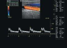
Recognized as the least costly option for vascular imaging, vascular ultrasound is common, and very effective at imaging vessels close to the skin surface. It is also noninvasive like CTA and MRA, but with ultrasound, there is no contrast material used and no radiation dose is administered. Ultrasound images are captured in real time, so radiologists can see the blood flow to organs and tissue in the body. Another notable benefit is the ability to do the procedure at a patient’s bedside. This can be significant if a patient is unable to be moved into a vascular lab for a CT or MR study.
Despite the fact that vascular ultrasound is very patient friendly, there are some deficiencies. Operator dependence is viewed by some as a problem. The dispute lies in the amount of time needed to train an individual on ultrasound. It takes longer to train to use ultrasound than a CTA or MRA, and the same results can often be viewed in much less time. Vascular ultrasound also has problems imaging vessels deep in the body, which can affect viewing vessels in bariatric patients.
Vascular diseases can be very deceiving because there are rarely symptoms in the early stages. However, advancements in vascular imaging technology will continue to enhance images, allowing for greater visibility of vessels, earlier detection and more accurate diagnosis of disease and hopefully more lives saved.
1. (Kock et al. Radiology 2005, Ofer et al. AJR 2003 and Rubin et al. Radiology 2001)


 March 05, 2026