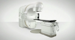
November 23, 2011 – The University of Alabama at Birmingham (UAB) Health System became the first medical center in the world last week to utilize intrafraction motion review (IMR), or "triggered imaging," to continually monitor tumor location during radiosurgery for lung cancer. IMR, which is a unique capability of the TrueBeam linear accelerator from Varian Medical Systems enables visual verification that a tumor is being properly targeted.
"With triggered imaging, clinicians use the imager on the TrueBeam system to observe the targeted tumor repeatedly, at a predetermined portion of the respiratory cycle, in order to check on the tumor's location and trajectory," said Chris Toth, senior director of marketing at Varian. "If the tumor is not where it is supposed to be, they can halt treatment and intervene to enhance the accuracy of the targeting."
Doctors at UAB used the IMR tool for the first time earlier this month when delivering a Gated RapidArc radiosurgery treatment for inoperable early-stage lung cancer. RapidArc enables fast, precise image-guided intensity-modulated radiotherapy (IMRT) by delivering dose continuously as the treatment machine rotates around the patient. Gated RapidArc makes it possible to monitor patient breathing and compensate for tumor motion during a RapidArc treatment. The gating system turns the treatment beam on and off in synchrony with the patient's breathing to increase treatment precision. With IMR, the gating system also triggers the imager to generate a low-dose X-ray of the targeted tumor at a specific point in the patient's respiratory cycle.
"With IMR, we can now monitor the gating accuracy and ensure that the beam hits the tumor," said Richard Popple, Ph.D., associate professor of medical physics at UAB. "It will allow us to monitor tumor position in real time and intervene if a change in patient respiratory pattern causes a shift. The enhanced precision could potentially increase tumor control and decrease the amount of surrounding healthy lung tissues exposed to the beam."
"The IMR tool offers us a way of verifying that our gating strategy remains valid throughout an entire treatment," said Chris Dobelbower, M.D., Ph.D., radiation oncologist at UAB. "It also gives us the ability to hold the beam should the target wander from isocenter if the patient's breathing pattern were to change due to a cough or a sneeze, or other interference. Those events did not happen with our first patient, so no treatment interruptions were necessary."
To utilize IMR during the treatment for lung cancer, Dobelbower collaborated with thoracic surgeon Douglas J. Minnich, M.D., assistant professor at UAB Healthcare, who placed a set of radio-opaque fiducial markers into the lung tumor using electromagnetic navigational bronchoscopy—an approach that employs three-dimensional computed tomography (CT) imaging to guide the procedure. These markers made it possible to see the tumor's location within the surrounding healthy tissues using X-ray images generated during treatment.
"IMR makes it possible to complete lung cancer treatments with a high level of precision and confidence that you're treating the area you want to treat," Minnich said. "Rather than imaging at the beginning of treatment and then doing your best to account for respiratory motion, IMR allows you to actually watch the tumor and monitor your targeting as the treatment proceeds."
According to Dobelbower, the ability to generate an image with every breath, identify when a targeted tumor has shifted, stop a treatment, and reposition the patient makes a new level of precision feasible. "This was previously only possible by a time-consuming and cumbersome process of interrupting treatment for additional imaging," he said.
For more information: www.varianmedicalsystems.com

 May 06, 2026


