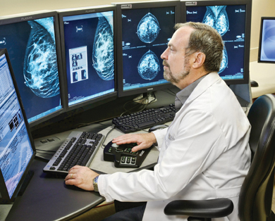
Radiologist Stephen L. Rose, M.D., knows that experienced breast radiologists paired with state-of-the-art equipment can make a life-saving difference in the early detection and diagnosis of breast cancer. A board-certified breast radiologist, Dr. Rose has been a big believer in the potential for tomosynthesis technology for more than five years. In 2009, Dr. Rose worked with the Bobetta Lindig Breast Care Center at Memorial Hermann Memorial City Medical Center in Houston, Texas, to install Hologic’s Selenia Dimensions digital tomosynthesis system and join the national clinical trial to evaluate this leading-edge technology. “I think tomosynthesis is going to revolutionize the way we screen for breast cancer,” declares Dr. Rose.
“The quality of images with Hologic’s two-dimensional digital mammography systems is excellent, but the ability to peel away layers with tomosynthesis is a tremendous advance and provides an opportunity to markedly improve what we’re doing in breast screening. Tomosynthesis makes a huge difference in the end result,” explains Dr. Rose.
Digital tomosynthesis is a three-dimensional imaging technology that enables radiologists to see “inside” the breast. With Hologic’s Dimensions tomosynthesis system, an X-ray tube rotates around the compressed breast taking 15 images from numerous angles, reducing interference caused by overlapping tissue that can obscure views and hide potential cancers.
Dr. Rose chose Hologic’s Selenia Dimensions 3D system because of its unique implementation of tomosynthesis technology. “I see Hologic as the leader in tomosynthesis,” states Dr. Rose. “I thoroughly researched the technology prior to recommending Hologic’s Dimensions 3D system; talking to other radiologists and manufacturers. I really like Hologic’s approach to tomosynthesis – combining 2D and 3D imaging on a single system, which gives us the flexibility to use both 2D and 3D imaging modes. No other vendor offers this combination in one system.”
Dedicated Breast Imaging Specialists Coupled with State-of-the-Art Technology Reduce Callbacks
Dr. Rose is president and founder of Rose Imaging Specialists, the largest community-based breast imaging practice in Houston, Texas. For the past 20 years, he has dedicated his radiology practice to the needs of breast patients, leading the way with full-field digital mammography, stereotactic biopsies, breast ultrasounds and breast MRI. The practice’s 13 breast radiologists serve eight major hospitals in the Houston area, providing women with a full range of imaging services. “We have helped revolutionize the way breast imaging is done in Houston,’’ says Dr. Rose. “We interpret between 65,000 and 75,000 mammograms annually, and every mammogram is interpreted by a breast imaging specialist.”
In addition to the Hologic Dimensions 3D system, Dr. Rose and the eight hospitals he serves transitioned to 16 Hologic Selenia Dimensions 2D systems, which are configured to convert easily to 3D breast imaging. “At the end of the day, I don’t think the cost of tomosynthesis will be all that different from digital mammography, yet it will enable us to reduce our callback rate significantly and increase our ability to find smaller breast cancers. So it really does appear to be a win-win situation, which very few new technologies provide.”
Dr. Rose considers a screening mammogram the most critical tool for the early identification of breast cancer. “If we don’t see anything on the screening mammogram, then it doesn’t matter what other tools or modalities we have because we won’t look any further. I think tomosynthesis is the biggest thing to happen to breast imaging in a long time. It is much more powerful, and it is a practical solution because unlike some modalities, tomosynthesis is easy to use and efficient, enabling us to screen almost everyone,” declares Dr. Rose.
Tomosynthesis Greatly Improves Specificity
“Tomosynthesis allows us to look at slabs to see if anything is hiding in the breasts,” continues Rose. “It allows us to be much more confident that an area is okay. We can see more clearly those areas we might have questioned on a digital mammogram and put to rest our concerns on questionable areas that we previously banged our heads against the wall trying to decide if they were something or not. With tomosynthesis it is pretty obvious, as we page through the area, if what we see is just breast tissue or something more significant. Tomosynthesis also will be a big help with cysts because it allows us to see the margins of the cysts and put concerns about those to rest as well.”
Initially, Dr. Rose looked to tomosynthesis as a tool for women with dense breast tissue, but he also has found it to be a valuable screening tool for women with fatty breast tissue. “I had a case recently where I did a digital mammogram and was concerned about an area, so I brought the patient back and one of my partners did an ultrasound. The patient also agreed to a tomosynthesis mammogram, which showed clearly that the area was cancer. This is a situation where we saw something with digital mammography, but we weren’t sure what we were seeing. Using tomosynthesis it was very obvious we were looking at cancer. So even with fatty tissue, there are cases where we will find very small tumors that otherwise would have been missed.”
As he looks to the future, Dr. Rose sees a real benefit to having both 2D and 3D imaging functionality on a single mammography system. “There are some cases where you might need to take a two-dimensional mammogram, such as patients with large breasts or with implants. I like having the capability to do both two- and three-dimensional imaging with all my systems, so tomosynthesis is available for every patient possible.
“Tomosynthesis is the most exciting tool I’ve seen in my professional career,” concludes Dr. Rose. “This technology will help us find cancers earlier with fewer callbacks for additional studies.”
Case study supplied by Hologic Inc.
The comments included in this article are the opinions and personal stories of the individual hospital and breast center and not necessarily those of Hologic.
Hologic, Dimensions and Selenia are trademarks and/or registered trademarks of Hologic Inc. and/or its subsidiaries in the United States and/or other countries.

 July 07, 2026


