Siemens Healthcare's MAGNETOM Verio is a 3 Tesla (3T) MRI system with a 70 cm open bore and total imaging matrix (Tim) technology, a system designed to deliver high-field imaging to patients who are claustrophobic, in pain or discomfort or those who weigh up to 550 lbs.
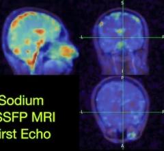
As a 3T MRI, Verio can be used for many applications, including neurology and functional neuro evaluation, orthopedic and cartilage assessment, breast, vascular and cardiac imaging. The 3T system reportedly optimizes parallel imaging, accelerates scan time and improves visualization as well as patient throughput.
The Verio is designed for treating obese patients by offering a bore that is larger than many conventional systems and a 550-lb table-weight capacity. The unit has a short length of any 3T system along with a 70-cm bore, which, according to the manufacturer, enables clinicians to expand care to otherwise hard-to-image patients such as children, patients challenged because of pain and mobility and the elderly. The larger space will also help when imaging claustrophobic patients because fewer patients will need sedation or refuse MR imaging services. Patient access is enhanced for both intensive care patients and MRI-guided interventional procedures and kinematic studies can be performed easier.
The Verio, with the power of Tim, has up to 102 seamlessly integrated matrix coil elements and up to 32 independent radiofrequency channels, enabling advanced clinical applications. Tim allows combinations of up to four different coils that make patient and reduces coil repositioning.
The syngo MR user interface, enables clinicians to evaluate pathologies, and are available on a 3T system with an open bore. Applications include: imaging in neurological, orthopedic and abdominal procedures, correcting for motion artifacts in reportedly all body regions; 3D MR angiography (MRA), obtaining arterial and venous phases in one scan and providing robust bilateral MRA, even in cases of a severe stenosis, says the manufacturer; obtains 3D imaging in all contrasts; for functional MRI; susceptibility weighted imaging to visualize blood products, allowing for better depiction of stroke and brain trauma patients, contusions and shearing injuries, and identification of intracranial vascular malformations.


 February 13, 2026


