May 11, 2012 - X-ray mammography is an important diagnostic tool in the fight against breast cancer, but it has certain drawbacks that limit its effectiveness. For example, it can give in false positive and negative results; it also exposes women to low doses of ionizing radiation, which – while accepted as safe – can still carry some risk.
In the first phase of clinical testing of a new imaging device, researchers from Netherlands’ University of Twente and Medisch Spectrum Twente Hospital in Oldenzaal used photoacoustics – light-induced sound – rather than ionizing radiation to detect and visualize breast tumors. The team's preliminary results, which were conducted on 12 patients with diagnosed malignancies and reported in the Optical Society’s (OSA) open-access journal Optics Express, provide proof-of-concept support that the technology can distinguish malignant tissue by providing high-contrast images of tumors.
"While we're very early in the development of this new technology, it is promising. Our hope is that these early results will one day lead to the development of a safe, comfortable and accurate alternative or adjunct to conventional techniques for detecting breast tumors," explained researcher Michelle Heijblom, a Ph.D. student at the University of Twente.
Photoacoustics, a hybrid optical and acoustical imaging technique, builds on the established technology of using red and infrared light to image tissue and detect tumors. This technology, called optical mammography, reveals malignancies because blood hemoglobin readily absorbs the longer, redder wavelengths of light, which reveals a clear contrast between blood-vessel dense tumor areas and normal vessel environments. However, it is difficult to target the specific area to be imaged with this approach.
As a means of improving this, the researchers combined the light-based system's ability to distinguish between benign and malignant tissue with ultrasound to achieve superior targeting ability. The result of their refinements is a specialized instrument, the Twente Photoacoustic Mammoscope (PAM), which was first tested in 2007.
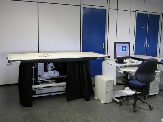
The device is built into a hospital bed, where the patient lies prone and positions her breast for imaging. Laser light at a wavelength of 1,064 nanometers scans the breast. Because there is increased absorption of the light in malignant tissue the temperature slightly increases. With the rise in temperature, thermal expansion creates a pressure wave, which is detected by an ultrasound detector placed on one side of the breast. The resulting photoacoustic signals are then processed by the PAM system and reconstructed into images. These images reveal abnormal areas of high intensity (tumor tissue) as compared to areas of low intensity (benign tissue). This is one of the first times that the technique has been tested on breast cancer patients.
By comparing the photoacoustic data with conventional diagnostic X-rays, ultrasound imaging, MRI, and tissue exams, the researchers showed that malignancies produced a distinct photoacoustic signal that is potentially clinically useful for making a diagnosis of breast cancer. The team also observed that the photoacoustic contrast of the malignant tissue is higher than the contrast provided by the conventional X-ray mammographies.
In looking to the future, notes Heijblom, "PAM needs some technical improvements before it is a really valuable clinical tool for diagnosis or treatment of breast cancer. Our next step is to make those improvements and then evaluate less obvious potential tumors, benign lesions, and normal breasts with it."
Paper: “Visualizing breast cancer using the Twente Photoacoustic Mammoscope: What do we learn from twelve new patient measurements?” Optics Express, Vol. 20, Issue 11, pp. 11582-11592 (2012).
For more information: http://www.OpticsInfoBase.org/OE

 June 02, 2026


