July 14, 2021 — RSIP Vision, a clinically proven leader in real-world AI solutions for medical image analysis, announced a new articular cartilage segmentation tool for accurate, non-invasive assessment of chondral lesions in MRI scans. This new software module enables quick and accurate deep learning-based segmentation of joint cartilage from MRI scans of hips, knees, and ankles. RSIP Vision’s advanced algorithms provide accurate measurement of the location and geometry of osteochondral lesions in MRI images that enable the physician to easily evaluate the extent of the damage, select the appropriate treatment and assess its efficacy.
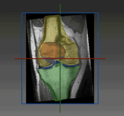
Chondral lesions are particularly prevalent among young and active patients including athletes. Due to the avascular nature of articular cartilage, the healing potential is limited. These lesions hinder the indispensable role that cartilage plays in creating a smooth, low-friction surface for transmission of forces across the joint. In many cases, Chondral lesions limit the athlete’s ability to participate in sports and even affect their daily activities. Cartilage segmentation is a crucial tool that aids the physician in choosing the optimal treatment for the patient. To help the physician assess the most suitable procedure from a variety of cartilage-sparing procedures (mosaicplasty, microfracture, osteochondral autograft transfer system, autologous chondrocyte implantation), the articular cartilage is imaged using MRI, segmented and then evaluated for lesion size (diameter), shape and boundaries. Any viable cartilage in non-weight bearing areas is also mapped for possible cartilage transfer (such as mosaicplasty or OATS).
RSIP Vision has extensive experience and expertise in MRI segmentation of soft tissues, offering fully automated segmentations with a submillimeter accuracy. The new module from RSIP Vision utilizes state-of-the-art deep learning (DL) algorithms designed to deal with MRI scans of cartilage in hip, knee, and ankle joints. The new technology is vendor-neutral and will be available to third-party MRI manufacturers and medical device vendors, allowing the physicians using the system, an improved way to evaluate cartilage damage, select the optimal treatment option and follow-up postoperatively.
RSIP Vision’s new module is essential in sports medicine, where cartilage damage is a common and debilitating problem among athletes from all fields. Using RSIP Vision’s module, physicians can provide athletes with better evaluation of their cartilage integrity and provide the best fitting cartilage-sparing treatment, resulting in improved outcome and shortened recuperation downtimes.
“It is very valuable to be able to accurately map chondral lesions preoperatively,” said Shai Factor, M.D., an orthopedic surgeon at Tel Aviv Sourasky Medical Center in Israel. “Analyzing the parameters of the lesion and its boundaries allows the surgeon, along with the patient, to choose the ideal cartilage repair technique. Additionally, in cases where cartilage transfer is the chosen option, this technology will make it possible to map the donor cartilage area as well and plan the surgery in the best way that will lead to better outcomes.”
For more information: www.rsipvision.com

 July 10, 2026


