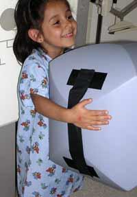
March 21, 2011 - A new positioning aid designed for pediatric patients undergoing chest X-ray exams, the Browning Ball by Supertech Inc. is supposed to make exams less fearful and the child more cooperative with the technologist.
Designed by Bruce K. Browning, RT, a pediatric radiographer at Kaiser Permanente, Southern California Medical Group, in Panorama City, Calif., the Browning Ball is a dual-technology aid. While playing with the ball prior to the exam helps the child feel less fearful and more cooperative with the technologist, hugging the ball for comfort during the exam also draws the scapulae out of the lung field.
A specialty in itself, pediatric radiology often requires a patient and considerate approach in order to obtain a high-quality diagnostic image – an attitude that is not always available at high-traffic imaging centers. One particular challenge in pediatric radiography is the anteroposterior (AP) projection of the chest on small patients with scapulae removed.
While there are several methods available for removing the scapula from the lung field – such as raising the arms above the patient’s head or having the patient positioned sitting or supine with the arms raised – these do not address the fears, frustrations and impatience of pediatric patients removed from their parents and surrounded by foreign diagnostic equipment. An alternative approach to imaging these sometimes-difficult patients is the Browning Ball.
“The ball is used to completely clear the lung field of the scapula, and this is done every time it is used,” Browning said. “Adults do this by hugging the upright chest unit or putting the shoulders on the chest Bucky. The foam of the Browning Ball is medical-grade and light enough for the majority of children to handle easily, without leaving artifacts in the images. Because the Browning Ball is made of foam or sponge and is essentially the same density as air, no change in exposure factors is necessary, and there is no increase in patient dose. The ball is also vinyl-coated for durability and easy cleaning.”
When using the ball, radiology techs should select an appropriately sized cassette and place it lengthwise with the patient at either end of the X-ray table, and then place the patient’s back against the cassette for the AP projection. The patient should hug the ball against the chest with the chin on top of the ball and hands out of the primary beam. The standing wall Bucky can then be lowered and adjusted for patients of different heights.
For more information: www.supertechx-ray.com

 January 22, 2026
