August 30, 2011 — Today's state-of-the-art diagnostics in digestive tract imaging, including capsule endoscopy, is the topic at MEDICA 2011, the world’s largest medical trade fair to be held in Düsseldorf, Germany Nov. 16-19, 2011. More than 4,500 exhibitors from 60 countries will be present.
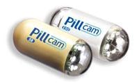
Capsule endoscopy can be used in all diseases of the small intestine in which conventional methods are unable or insufficient to shed any light. Instead of a relatively thick endoscope, patients now only need to swallow a very small capsule containing two tiny cameras that provide clear images of the interior of the stomach and intestines.
The process is not new. However, technical advances now even make it possible to move the capsule where it is supposed to go – controlled and manipulated by magnetic force. A particularly sophisticated model is a capsule that moves on its own "two feet.”
Private lecturer Jutta Keller, M.D., of the medical clinic at the Israeli Hospital in Hamburg, says finding the sources of bleeding in the small intestine remains the most important indication for the method.
Based on a resolution of the Joint Federal Commission of 2010, with this indication capsule endoscopy is now paid for by the German healthcare system. The method is recommended and applied, amongst others, in cases of suspected Crohn's disease in the small intestine, complicated sprue and when other tumors of the small intestine are suspected.
In addition, says Keller, there are systems for examining the esophagus and colon, the limits of whose clinical uses are not yet clear. Capsule diagnostics is not indicated for passage disorders of the gastrointestinal tract. It should also not be used on patients with pacemakers, those with a history of multiple abdominal operations, and pregnant women.
The method cannot yet completely replace conventional endoscopy of the stomach and colon. One reason is that biopsies are not possible. According to the gastroenterologist, one of the greatest disadvantages of capsule endoscopy to date has been the lack of control over the capsules. For that reason, new and different systems have been or are being developed:
Guiding hand-held magnets
The most advanced systems are controlled by means of magnetic forces. This requires a capsule made of magnetic material and an exterior magnetic field. One option, for example, is control by means of a hand-held magnet. Such a system was developed in a European research project (NEMO: nano-based capsule endoscopy with molecular imaging and optical biopsy). According to Keller, the system is based on a hand-held magnet developed by the Fraunhofer Institute in St. Ingbert and a modified capsule endoscope developed by the Israeli company Given Imaging Ltd. He says that initial clinical trials with the system were quite successful. A targeted and comprehensive examination of specific structures was successful in most subjects, in that most of the mucosal lining of the stomach could be displayed.
Electromagnetic maneuvering
Siemens and Olympus have jointly developed a second system: MGCE – magnetically guided capsule endoscopy. The magnetic capsule (length 31 mm, diameter 11 mm) can be remotely navigated through the stomach by means of a joystick. The capsule with two cameras and a permanent magnet takes four pictures per second; they are then transferred wirelessly to an image processing system and can be viewed immediately.
The results of a first study were presented and verify that this method works. Of the more than 50 people examined, a total of 30 showed pathological changes in the stomach. Of those, 14 were discovered using the capsule as well as a conventional gastroscope, ten with the capsule only, and six with the gastroscope only.
"In gastrointestinal examinations, the magnetically controlled capsule produces results that are as reliable as those obtained by conventional endoscopy. However, stomach examinations using the capsule are more comfortable," concludes Studies Director Jean-Francois Rey, M.D.
Since the procedure requires practice, Siemens has developed a so-called stomach simulator. It contains an anatomically accurate model of the stomach. Every possible capsule maneuver is simulated. The simulator reproduces the video image of the capsule and displays the same navigation information available to doctors doing a real examination.
According to Keller, the results of current studies plus technical advances are "impressive" and promise clinical capsule endoscopy examination of the stomach will be possible in the future. For this and, examinations of the esophagus and the retroperitoneally fixed duodenum, the simple hand-held magnet system could be a "pragmatic, effective and inexpensive solution."
It appears to be less suitable for examination of intraperitoneal sections of the intestine; the one-sided magnetic field generated will tend to move the mobile loops of the small intestine instead of maneuvering the capsule longitudinally through the loops. By comparison, a precisely controlled electromagnetic system could maneuver a capsule to "float" through the entire gastrointestinal tract. However, the currently available system is "still a long way from achieving these requirements," explains the gastroenterologist from Hamburg.
On eight "feet" through the porcine intestine
Other developments are not yet ready for day-to-day use, says Keller. VECTOR (Versatile endoscopic capsule for gastrointestinal tumor recognition and therapy) is a "video capsule with feet", developed in a further European research project. The aim was a capsule allowing targeted movement through the stomach and intestines. Preliminary data show that an eight-legged capsule can "move through a phantom model of an ex vivo colon". In an in vivo porcine intestine, the capsule "automatically moved a limited distance (15 cm)." However, this model did not have any control mechanisms.
In addition, attempts are being made to maneuver capsule endoscopes through the gastrointestinal tract by means of electrical stimulation. Electric pulses emitted from the capsule to the gastrointestinal wall trigger a contraction of the muscles in front of or behind the capsule. In this way, the capsule is moved in an oral or aboral direction. Systems such as this could be especially suitable for examinations of the esophagus and small intestine.
The further technical development of capsule endoscopy is, of course, not limited to capsule navigation, emphasizes the Hamburg gastroenterologist Dr. Ingo Steinbrück. Battery power, visual range and image quality, amongst other things, were and are still being developed further. These developments aim to "enhance diagnostic sensitivity, optimize energy consumption, shorten the waiting time for findings and increase acceptance by patients." One of the most important objectives for gastroenterologists remains intestinal cancer screening at home.
MEDICA 2011 is not just showcasing state of the art gastrointestinal diagnostics and therapy in terms of imaging systems such as the respective endoscopes; the MEDICA Congress will include the event "Detection of early-stage lesions in the gastrointestinal tract" headed by Prof. Dieter Häussinger, M.D., on Nov. 17 at the Congress Center Düsseldorf (CCD.South).
For more information: www.medica-tradefair.com

 May 19, 2026