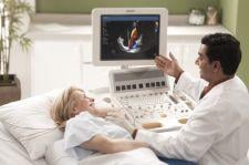
Philips has a new addition to its HD ultrasound family of products, the Philips HD15 ultrasound system, which is designed to provide physicians with high-end imaging and workflow performance in a cost-effective system.
The HD15 is a new platform designed to deliver an advanced level of image clarity and broad application support for everyday use in small hospitals, clinics and private practices. The system may be used as a primary system for some users, particularly those in emerging markets who require a feature-rich system but may not need all of the features of a high-end ultrasound solution. The HD15 contains multiple usability features to improve workflow, as well as versatile capabilities for a wide range of exam types including general imaging, cardiac, vascular and OB/GYN applications. In addition, advanced features like contrast enhanced ultrasound and PureWave transducer technology allow users to perform real-time guidance and evaluation of minimally-invasive treatment procedures and provide more diagnostic confidence on technically challenging patients and pathologies.
Philips technologies such as QLAB quantification software, XRES image processing and PureWave transducer crystal technology have been integrated into the HD15 to provide clinicians a robust system that assures ease of use and productivity. New Microfine EX focusing provides sharper images and improved tissue uniformity throughout the depth of field through application of new dynamic receive lens tuning with five times more focal points than previous generation systems. Tissue Specific Imaging presets and iSCAN one button image optimization can quickly provide clear images with little to no adjustment. A broad suite of configurable patient reports and exam storage options, such as DVD-CD-R/RW, USB drive, and full DICOM capabilities, provide efficient patient data management and colleague or specialist consulting.
The HD15 offers active native data to allow clinicians to manipulate exam parameters and image settings even after the patient has left. Images and Cineloops can receive further investigation by manipulating the original image to see new detail. Live compare reportedly lets the clinician compare a previous exam side-by-side with an active exam in order to immediately see changes in structure or blood flow. This can be particularly used in comparing changes in cardiac and vascular anomalies, further documenting changes after interventional procedures or evaluating fetal development.

 May 07, 2026

 Clearance")


