February 7, 2012 — CIRS is pleased to introduce a new cone beam computed tomography (CBCT) Electron Density and Image Quality Phantom. The modular system is a single tool configured to measure twelve performance parameters necessary for electron density calibration in volumetric imaging and image quality analysis on CT and CBCT imaging systems during commissioning and periodic quality assurance (QA).
The system’s ease of use allows CT/CBCT users to perform routine measurements for assessment of alignment, spatial uniformity, low contrast visibility, magnification/spatial linearity, CT number linearity, contrast-to-noise ratio (CNR), slice thickness, spatial resolution, modulation transfer function (MTF), noise, size independence, and absorbed dose.
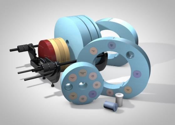
The system is divided into 3 modules that allow for multiple configurations to be purchased separately: Electron Density Phantom (062M), Electron Density Phantom (062MA) and CBCT Electron Density and Image Quality Phantom (062MQA).
The Electron Density Phantom (062M) provides a tool for CT number to electron density calibration. The CBCT Electron Density Phantom (062MA) accounts for the specific geometry of cone beam kV and MV CT imaging for axial/helical CT equipment due to the imaging volume that closely resembles an average male torso, and accommodates an ion chamber for dose measurements. The complete CBCT Electron Density and Image Quality Phantom system configuration (062MQA) is optimized for holding unit (HU) to electron density calibration in volumetric imaging, and includes an image quality tool specifically designed to perform image QA tests for CT as recommended in Report #1 of the AAPM Task Group. The phantom is also compliant with AAPM Task Group 142.
For more information: www.cirsinc.com

 June 18, 2026


