Sept. 8, 2026 – Brainomix and Endeavor BioMedicines recently presented positive results from the Brainomix AI-driven quantitative analysis of HRCT imaging data in Endeavor's Phase 2a trial of taladegib in idiopathic pulmonary fibrosis (IPF) at the European Respiratory Society (ERS) Conference in Barcelona.
The randomized, double-blind, placebo-controlled 12-week study evaluated taladegib, a hedgehog pathway inhibitor in patients with IPF. The trial previously demonstrated that treatment with taladegib improved lung function from baseline as measured by forced vital capacity (FVC), increased total lung capacity and reversed measures of interstitial lung disease (ILD) from baseline at Week 12.
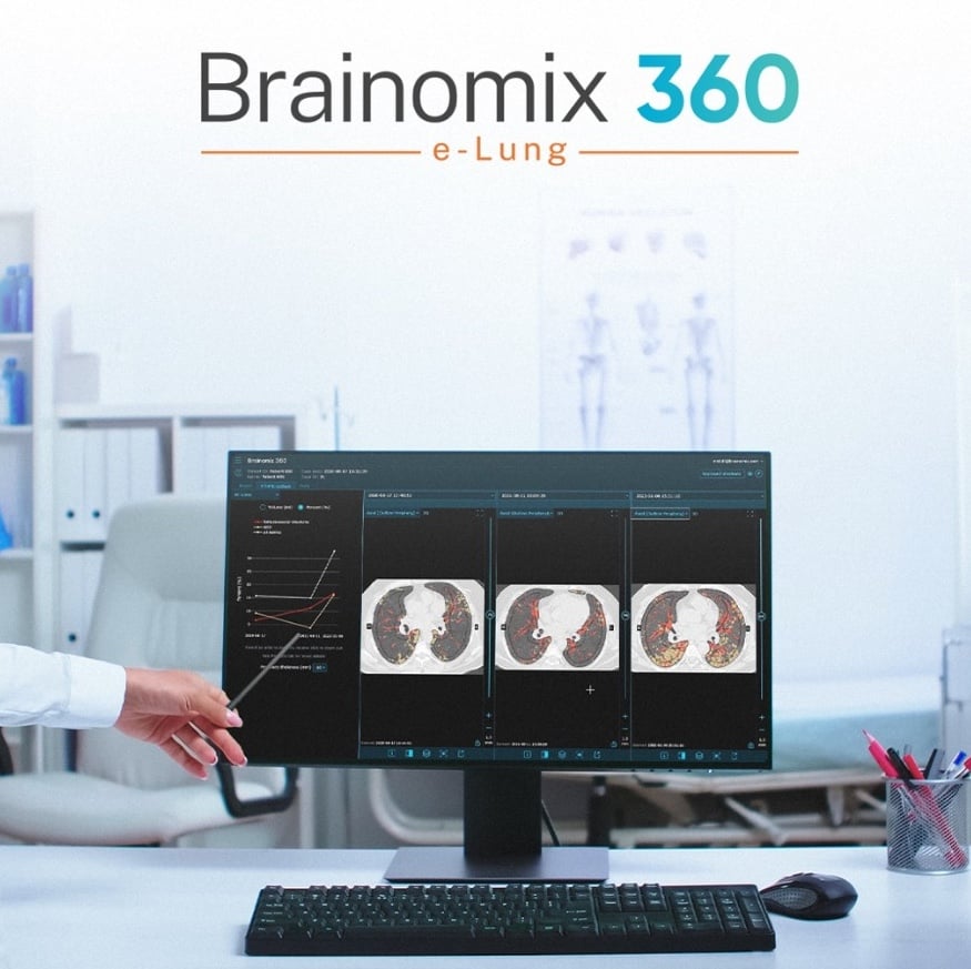
Brainomix 360 e-Lung is an FDA-cleared, CE-marked AI-powered image analysis tool that provides automatic quantifications and visualizations of radiographic features in lung CT scans. Clinicians can use these outputs to aid in their diagnostic assessment of lung disease.
Brainomix and Endeavor entered into a collaboration to conduct a post-hoc analysis evaluating the potential of Brainomix AI-powered quantitative imaging outputs to support clinical trials. Trained on diverse datasets, the platform has already been employed in several Phase 2 and Phase 3 trials and became the first quantitative CT imaging platform selected as a co-primary endpoint in a Phase 3b pulmonary fibrosis trial.
IPF Trial
n a post-hoc analysis of the 12-week Phase 2a ENV-IPF-101 trial, e-Lung quantitative CT imaging was used. Analysis by the authors detected significant positive differences in favor of taladegib in lung volume, overall ILD burden and total fibrosis extent.
These findings highlight the potential of AI-powered quantitative imaging outputs to support the detection of meaningful treatment effects in small, relatively short clinical studies, supporting more efficient and informed clinical development.
Importantly, these results highlight the value of reliable, objective AI-endpoints in clinical development programmes for pulmonary fibrosis. While forced vital capacity (FVC) remains an established clinical endpoint, it may not fully capture meaningful biological changes or provide mechanistic insights into treatment response. As background therapies reduce measurable declines in lung function, increasingly sensitive CT quantification results may help detect subtle treatment effects and support more efficient, data-informed drug development.
These positive results have catalyzed further collaboration between Brainomix and Endeavor BioMedicines, aiming to incorporate quantitative image analysis into the further clinical development of taladegib for pulmonary fibrosis.
Dr.Lisa Lancaster, Chief Medical Officer at Endeavor BioMedicines, commented: “The results of Brainomix’s post hoc quantitative imaging analysis continue to support taladegib’s potential to improve lung structure, reduce fibrosis and improve lung function in patients with IPF. We are impressed with the technology’s ability to detect statistically significant changes in a small cohort and over a short treatment duration. These results are a testament to the value of objective, sensitive and reliable endpoints for the de-risking of clinical development, and we look forward to future collaboration opportunities with Brainomix.”
Professor Peter George, Consultant Pulmonologist at the Royal Brompton Hospital, UK, and Senior Medical Director at Brainomix, added: “Our goal is to accelerate the clinical development of potentially transformational treatments, and taladegib is one of the most promising therapies for pulmonary fibrosis currently in development. We are very pleased to see that our analysis with e-Lung was able to provide key insights into the efficacy and mechanism of taladegib. Quantitative CT imaging offers a powerful approach to support earlier, faster, and more informed development decisions, ultimately helping promising therapies reach patients sooner.”

 October 07, 2026