
Dave Fornell is the editor of Diagnostic & Interventional Cardiology magazine and assistant editor for Imaging Technology News magazine.
Blog | Radiology Imaging | December 22, 2015 | Dave Fornell
X-rays and Mom — My Own Personal Case Study into the State of Imaging Technology

Figure 1. A bedside screen shot of a Carestream DRX mobile X-ray in the ED of the fractured fibula.
While I write a lot about medical imaging technology and how new technology can and should work, it is not often that I get to experience how things actually work in the real world. This past Thanksgiving I received a call from a paramedic explaining that my mom had fractured her leg and I should stop working on the turkey and fixings and rush to the emergency department (ED) at Edward Hospital in Naperville, Ill. She had been walking her dog on wet grass and leaves in a park when her dog took off after another dog and pulled her down. She was whipped around and the change in weight caused her to dislocate her ankle (the bottom of her shoe was facing her when she looked at her feet) and caused a spiral fracture to her fibula.
When I got there my mom was already heavily sedated due to the pain and because the ED staff had already put her ankle back in place. The ED doctor ordered a digital radiograph (DR) of her leg to see the extent of damage. They wheeled in a new Carestream DRX mobile X-ray system and I had a live demonstration of how fast these types of systems can snap the pictures. It called up the images immediately on the machine’s screen. The image of the Pott’s fracture with fragments (figure 1) was really interesting as someone who covers radiology, but I also realized from a non-clinical standpoint she was really messed up and in pain. Additionally, she would need reconstructive surgery to put her Humpty Dumpty leg back together again. She was way up the creek without a paddle with it being Thanksgiving and there were no orthopedic surgeons in staff due to the holiday. The day after Thanksgiving was not much better, as we found, since most physicians were out through the following Monday. So the ER splinted the leg, wrapped it in ace bandages and sent her home with heavy pain killers.
Compounding her mobility issues was the fact that she has bilateral knee replacements. (Figure 2) Due to the trauma, broken bone and knowing she had these implants that further limited her ability to move around, she was prescribed a prophylactic anticoagulant.
Knowing we would need the images for a surgeon to review, I had the ED burn a CD. However, I was happy to find Edward is among the growing number of hospitals to grant patients access to their health records via a DR Systems Internet image/results distribution system. This technology pulls images and reports from the hospitals’ Epic EMR (electronic medical record) system and makes them available for remote viewing by clinicians outside of the hospital’s picture archiving and communication system (PACS). She also was given login instructions at discharge for a patient portal so she could access her records and images herself on a home computer or smartphone.
We managed to find one orthopedic surgeon in their office on the Friday after Thanksgiving. They thought it was great that we had a CD, but before attempting to open it, they asked which hospital she had been at. Edward was already in a health information exchange, so outlying offices such as this one from a different medical group could access her records remotely in less than a minute. (Figure 3) They were able to call up her images and see what meds she was prescribed, which made the office visit go much faster.
She had surgery on Dec. 1, the Tuesday of RSNA 2015. The orthopedic surgeon practiced at Elmhurst Hospital in Elmhurst, Ill., across the county from Naperville. But, thanks to the remote image viewing system, they could get the ED images for reference and planning. The surgeon’s post-surgery DR image showing the reconstruction of the fibula also was available via my mom’s patient portal. (Figure 4)
She did what most patients today do with this type of access and posted her X-rays on Facebook. Leveraging the Facebook form of patient engagement, the result was lots of sympathy, flowers and friends volunteering to help her with things around the house and groceries since she cannot walk or drive for at least two months.
While an unfortunate incident and a horrible thing to have happen to my mom, from a professional standpoint, I was happy to see the technology I cover working in the real world as it was intended. The speed in workflow efficiency, speed and ease of access to her imaging at the point of care and remotely, and access to a patient portal are all examples of how the healthcare system should work. In this case, the technology and imaging integration was flawless.
Find other radiology insights in ITN's blog section.
-
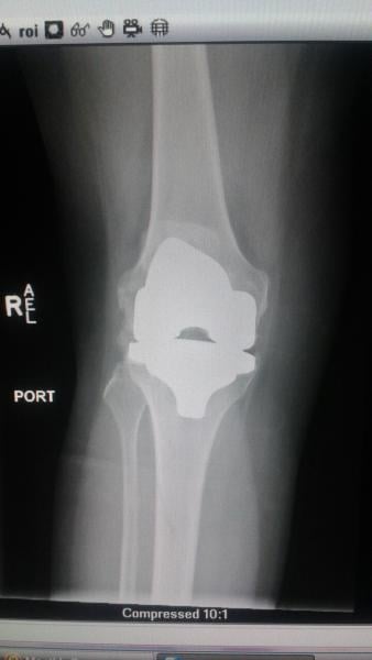
Figure 2. Another ED bedside DR image to confirm there was no secondary fracture at the top of the fibula. The image shows one of my mom's knee replacements.
-
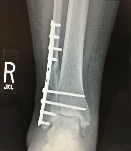
Figure 4. The post-surgical X-ray showing the bone repair, which was accessed and copied by the patient using a patient portal.
-

Figure 3. Across the county from the ED, an orthopedic surgeon's office was able to quickly pull up her X-rays using a remote image viewing system that granted access to the patient's EMR information, reports and medical imaging. The little girl is my daughter Vivian playing doctor with grandma to keep her spirits up.
Related Content

News | Pediatric Imaging
June 16, 2026 — Crescom has officially launched a global clinical Proof of Concept (PoC) of its pediatric ...
 June 24, 2026
Feature | X-Ray | Kyle Hardner
Water-window X-rays allow researchers to visualize biological cells at high contrast without staining agents or other ...
June 23, 2026 
News | Artificial Intelligence
June 15, 2026 — HOPPR recently announced that HOPPR AI Foundry is now available in AWS Marketplace. The availability ...
June 19, 2026 
News | Radiology Imaging
June 15, 2026 — Lead Glass Pro, a supplier of radiation shielding products, has expanded its turnkey installation ...
June 18, 2026 
News | Digital Pathology
June 15, 2026 — Leica Biosystems is expanding the availability of its Aperio GT Elite digital scanner into the EMEA ...
June 15, 2026 
News | Radiology Business
June 9, 2026 — Bayer has appointed Dr. Jost Reinhard president of the Radiology business within Bayer’s Pharmaceuticals ...
June 12, 2026 
News | Enterprise Imaging
June 9, 2026 — GE HealthCare will showcase its latest enterprise imaging solutions at the Society for Imaging ...
June 09, 2026 
News | Digital Radiography (DR)
June 3, 2026 – Konica Minolta Healthcare Americas has announced a new version of AeroRemote Insights, the company’s ...
June 05, 2026 
News | Innovative Hospitals
May 27, 2026 — Nearly two years after announcing plans for a “real-world” academic-industrial collaboration, GE ...
June 03, 2026 
News | Radiology Business
May 22, 2026 — The American College of Radiology (ACR) supports passage of the Medicare Access to Radiology Care Act (S ...
May 26, 2026 © Copyright Wainscot Media. All Rights Reserved.
Subscribe Now