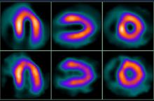
Stress images: top row, images acquired with quarter-time protocol and processed by WBR bottom row, images acquired with full-time protocol and processed with FBP, with all other acquisition and dose parameters identical.
March 2, 2009 - Results were released today from a clinical study of the quarter-time Xpress3.Cardiac, UltraSPECT's WBR (wide-beam reconstruction) image reconstruction algorithm used to create half-time SPECT images of high diagnostic quality.
The study was conducted in the Division of Nuclear Medicine, Department of Radiology, St.-Luke’s-Roosevelt Hospital in New York It was conducted by a group of researchers led by Dr. DePuey, M.D., director of nuclear medicine at St. Luke’s-Roosevelt and professor of radiology at Columbia University.
“For perfusion SPECT, quarter-time WBR affords image quality, defect characterization, and functional assessment equivalent to full-time OSEM, providing the potential for decreased SPECT acquisition times and/or decreased radiopharmaceutical doses,” said Dr. DePuey. “However, users should be aware of the differences between the various methods when reporting functional results; compared to full-time gated SPECT, quarter-time gated WBR yields significantly lower LVEF's.”
Since the introduction of the half-time Xpress.Cardiac in 2005, UltraSPECT's WBR image reconstruction algorithm has been in routine clinical use for nuclear cardiology, producing half-time SPECT images of high diagnostic quality. WBR incorporates simultaneous resolution recovery and noise control during reconstruction without applying a post-processing filter. The purpose of the current study was to clinically evaluate the more recently introduced Xpress3.Cardiac product for quarter-time imaging, and determine whether this image acquisition protocol processed by the WBR algorithm optimized for quarter-time SPECT produced adequate imaging outcomes.
The study used the optimized algorithm, in which 209 patients (91 men, 118 women, mean chest circumference = 40 inches) were imaged at rest and stress (dosage: 9 and 32 mCi99 Tc-sestamibi, respectively), first full-time using OSEM (and FBP), and then quarter-time with the new optimized WBR algorithm. Full-time rest scans were acquired in just over 14 minutes (180 degree arc covered by 64 stops at 25 seconds per stop (sps) with a 90 degree, angled, dual-headed detector), while full-time gated (eight frames per cardiac cycle) post-stress scans were performed at 20 sps. Each of these tests were immediately followed by their quarter-time counterparts: rest (6 sps) and post-stress (4 sps) gated SPECT. Blinded observers graded scan quality (1 = poor to 5 = excellent) based on myocardial uniformity, endocardial/epicardial edge definition, and background noise. Perfusion defects were scored using a 17-segment model. In addition, three different commercially available software methods were used to calculate EDV, ESV, and LVEF, Dr. DePuey said.
The results of the study showed that for rest studies, the mean image quality for quarter-time WBR was equivalent to those of full-time OSEM and full-time FBP. For stress, quarter-time WBR quality was actually superior to full-time OSEM. In patients with chest circumferences greater than 44 inches, a longer, 10 sps WBR acquisition improved resting image quality, researchers said. Mean SSSs, SRSs, and SDSs were not significantly different with quarter-time WBR vs. full-time OSEM or FBP.
As for the quantitative analysis of LV volume and function, all three software methods employed showed a good correlation between LVEF, EDV, and ESV as determined by WBR vs. OSEM, although ESV's were generally higher with WBR, primarily due to better delineation of the valve plane at end-systole; EDV's, however, were similar. Thus, EF's were significantly lower with WBR, the study concluded.
For more information visit our website: www.ultraspect.com

 June 15, 2026


