Varian's Trilogy linear accelerator, fitted with OnBoard Imager, gives doctors the ability to track and target tumors.
Many patients who were previously turned down as surgery candidates because their tumors were considered unreachable are getting a second chance.
Extremely accurate dose delivery though stereotactic body radiotherapy (SBRT) is enabling clinicians to reach formerly unreachable tumors. However, it has been limited to areas of the head and neck until quite recently. Through cone-beam CT, SBRT can now be used to target tumors throughout the body.
Because SBRT delivers dose more precisely than other techniques, like IMRT and IGRT, it causes less damage to surrounding tissue. Its accuracy also enables more dose to be delivered at each treatment, thus reducing the number of sessions a patient must attend.
The challenge with SBRT is that it was previously not feasible for body components that are nonrigid and associated with substantial organ motion. Now, thanks to cone-beam CT, SBRT is being used in many other areas of the body, including the abdomen, which experiences considerable movement due to breathing as well as stomach, bladder and rectal filling.
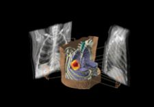
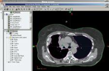
With the incorporation of cone-beam CT and SBRT, doctors are able to immediately compare 3D axial CT slices with initial treatment planning images to determine the exact location of a tumor prior to each treatment. This also facilitates the delivery of a higher dose.
Cone-Beam CT Allows for Tighter Margins
When using cone-beam CT to verify an accurate treatment plan for SBRT, cone-beam images are directly overlaid onto original planning images, allowing physicians to compare the position of the tumor in relation to various structures surrounding it.
“Cone-beam CT offers the direct, actual, right-at-the-time-of-treatment look at how the organs are positioned and how the dose will fall relative to them, as well as a comparison to what was planned for the ideal situation for that patient. If there are discrepancies, we can, in real-time, make changes before treatment,” said James McGee, M.D., S.M, of OSF Healthcare in Peoria, IL.
According to Dr. McGee, it is important to reduce the margins in order to spare normal tissue when delivering radiation dose. However, reducing margins without verifying where the tumor currently is positioned increases the chance that it will not be adequately treated.
“In the past, we have had to allow rather significant margins around the area being treated because we knew that the setup uncertainty was a very real part of daily management,” said Dr. McGee. “Now we accept the fact that there will be changes from day to day, but we know that with the [cone-beam CT] we can adjust the positioning of the radiation in order to take that into account each time.”
Dr. McGee uses Varian Medical Systems’ linear accelerator Trilogy, fitted with the On-Board Imager device. The Trilogy is optimized for both conventional and stereotactic approaches to treating cancer and can be used to deliver 3D conformal radiotherapy, IMRT, stereotactic radiosurgery, fractionated stereotactic radiation therapy and intensity-modulated radiosurgery. Its On-Board Imager accessory is designed to improve the precision and effectiveness of cancer treatments by giving doctors the ability to accurately target and track tumors safely, while the patient is in the treatment position.
According to John Buatti, M.D., of The University of Iowa Hospital, who uses Siemens Medical Solutions’ MVision Megavoltage Cone Beam (MVCB) Imaging Package, by monitoring intrafractional movement between one treatment day and the next, physicians are able to verify and retarget to the most accurate position possible.
Designed to work with Siemens’ linear accelerators, the MVCB utilizes a standard radiotherapy treatment beam. MVision makes it possible for the megavoltage source used for treatment to also create a 3D image of the patient, enabling clinicians to “see inside” the patient at the most appropriate moment.
“The best thing about the MVision is that it is readily able to be implemented and has been relatively quick from a daily treatment basis so that the actual implementation, delivery and execution has not been terribly time consuming or ornate to the point that it is extending treatment times,” said Dr. Buatti. “It’s very efficient.”
Before cone-beam CT Dr. Buatti used regular flat panels and films and a real-time optic-guided 3D ultrasound. “We’ve done some analysis and found that the image interpretation, speed and variability options are better with the cone beam,” he said.
Keeping an Eye on Tumor Movement
Though one of the major goals of radiation oncology is to spare as much healthy tissue as possible, patients undergoing extensive treatments, some of which may last weeks or months, physically change and so do their tumors.
When unchecked, these changes allow for damaging healthy, surrounding tissue with traditional radiation therapy, especially in tightly surrounded areas of the body and those that move significantly during breathing.
Dr. McGee uses the fluoroscopy component of the On-Board Imager in order to overcome motion when treating patients. “We know that the pancreas moves a great deal during breathing, as does the small bowel that is wrapped around it,” he said. “We fluoro and watch the movement of the pancreas with fiducial markers that are placed within it. The nice thing about cone-beam CT is that in many of these situations we are not just relying on markers. We are able to really see the whole volume and change relationships.”
Another feature of the Trilogy On-Board includes monitoring and gating for tumor motion. It characterizes respiratory motion and automatically uses the information to verify patient positioning and deliver treatments corrected for tumor motion. According to Dr. McGee, with fluoroscopy and respiratory gating, treatment accuracy can be as precise as one millimeter.
“We are actively using [cone-beam CT] in all of our head and neck patients and all of our prostate patients, but I think that there are a large number of other sites where it is going to prove itself extremely useful, particularly lung and abdominal tumors,” said Dr. Buatti. “I think it’s going to become a very commonly used technology for setups.”
Stronger, Fewer Sessions Equal Faster Recovery and Pain Relief
By verifying plans with cone-beam CT before each treatment, physicians are able to safely reduce margins and write prescriptions for higher doses. Additionally, by having higher doses delivered to more exact locations, the number of sessions required is reduced dramatically.
“Just by having the dose really on the target each day is a way of increasing the doses to the tumor because we are not having days where we partially missed the tumor,” said Dr. McGee.
By delivering stronger doses more precisely, patients are experiencing pain decrease in just days as opposed to weeks and weeks, according to Dr. McGee. In some cases, such as patients with lung cancer, only three treatments were needed, as opposed to 35.
“We have been able to treat patients with painful spine metastasis and cancers with a single treatment as opposed to 20 in the past,” he said. “The nice thing about that is the single high-dose fraction works much better, so not only are patients just coming in once instead of for four weeks, but within a few days their pain is getting much better as opposed to just a modest response.”
4D Dosimetry – The Next Step
With cone-beam CT leading the way in treatment verification, the next step is 4D dosimetry for radiation therapy. There is one stumbling block – dosimeter tools may not yet be up to the task of comprehensively verifying advanced
4D treatments.
“There is hope that ultimately we’ll be able to change the shape of a radiation field as we get [closer] to a treatment day or when the tumor has changed in shape, and be able to plan very quickly on the spot to make an adjustment for that change,” said Dr. McGee.

 June 18, 2026


