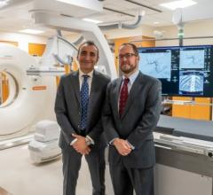
Doug Ryan, director, CT business unit, Toshiba America Medical System Inc., Tustin, CA
While much of the buzz in recent years surrounding computed tomography technology has focused on the escalating number of slices newer units are capable of delivering, there's more to the modality than just how to speed up the process.
Certainly, the additional slices are enabling CT to move into new imaging arenas, such as cardiology, neurology and trauma, giving clinicians the ability to scan virtually anything in less than a minute with remarkable clarity and detail. For example, CT units today allow clinicians to scan the heart and coronary arteries in five seconds or conduct a full-body scan in 30. Mobile units have been used to image mummified remains from Egyptian pyramids to authenticating Stradivarius violins.
But not everyone in the outpatient imaging community plans to - or even wants to - specialize in cardiology, neurology or trauma, and may not have the wherewithal to dabble in anthropology or musical history.
Many rely on their tried and true CT units as the capable workhorses that they are in their facilities. The single, four-, eight-, 16- and 32-slice models may seem like yesterday’s technology but they still have useful applications today. The question is whether they’ll be as useful tomorrow, particularly if 64-slice technology and higher sees their prices drop over time as demand and supply increases.
Executives from two of the leading CT manufacturers – Siemens Medical Solutions Inc. and Toshiba America Medical Systems Inc. agreed to share their forecasts and intelligence with Outpatient Care Technology on the modality's progression in outpatient care facilities.
What are some of the more recent noteworthy improvements in CT and where are the growth areas for CT applications today in the outpatient arena?
Doug Ryan, director, CT business unit, Toshiba America Medical Systems Inc., Tustin, CA
Utilization of CT has grown tremendously since inception and particularly in the last eight years since the launch of multislice technology. The major growth area has been in CT angiography, in addition to the patient throughput and image quality improvements in chest/abdomen/pelvis and neuro CT exams. The addition of advance visualization in CT angiography has been a tremendous benefit to the healthcare community resulting in fast and accurate diagnosis. This has been achieved by both technical and software innovations that have resulted in enhanced image quality and the ability to provide automation in complex scan procedures, which have significantly increased patient throughput and reduced the time required for diagnosis.
Scott Goodwin, vice president, CT division, Siemens Medical Solutions USA Inc., Malvern, PA
One of the more recent noteworthy improvements is the emergence of dual-source CT technology, which is our Definition product. It provides two sources and two detectors that can be run in real time using two different energy levels. Traditional CTs have one X-ray tube and one detector. Definitionss two X-ray tubes and two detectors allow you to look at things you never could before, such as tissue characterization, plaque quantification and the ability to produce noncontrast studies from a contrast study. You also have real-time bone subtraction where in CT today you have to manually edit that out. With the dual-source technology you can do it at the push of a button. Another benefit is the pure outright speed of the system, which has a temporal resolution speed of 83 milliseconds. Our 64-slice technology comes in at 165 milliseconds, while others do it in 175 milliseconds. This capability allows you to image the entire heart regardless of the heart rate. You don't need to use beta blockers to slow the heart rate down so that saves a lot of money. It reduces costs because you don't have to use beta blockers or use the nurse to administer them and you don't need to wait for the beta blockers to work in the patient. It gives you the ability to image faster.
The two X-ray sources also allow you to successfully image bariatric patients. The product delivers enough power to penetrate the necessary tissue and scan the bariatric patient without slowing scan speed. So it uses less power to image faster. The 64-slice scanners require you to slow the heart down to take the image. As a result, the diagnostic challenges are solved and replaced by clinical challenges. The question becomes how do you want to use it to diagnose the patient?
Another of the new improvements involves the size of data sets. As speed and resolution increases, data sets are getting bigger and bigger. When you look at the ever-growing data sets being developed and collected you have to determine what you want to do with them. How do you read them? Store them? Distribute them? To help clinicians in this process, we added computer-aided detection capabilities for the lung and the colon as an overlay.
One of the key challenges is how do you move these data sets around? We developed [syngo] WebSpace, a thin-client server to facilitate this process from a laptop or home or office PC. That way you don't have to be at a designated workstation to access data. You can do it remotely without having to go through a network so you don't have to invest in or go to any one of multiple workstations to accomplish your tasks. All you need is online access and multiple licenses. This is particularly helpful for clinicians who move from hospital to hospital regularly or that work in outpatient settings. You can tie your referral base into multiple systems and access it remotely via laptop and even do consults.
Each of the newer models of CTs - multislice (including the new 256-slice model), dual-source, cone-beam, helical and volumetric CT - offer a variety of different benefits to the clinical end-users and their patients. What are they? And how will reimbursement be adjusted - if at all - to accommodate for these higher-end CT technologies?
RYAN: The most important milestone for CT today is to reduce the radiation dose during an exam. The ability to scan an organ in a single rotation with a 256-slice CT scanner provides this benefit by eliminating the need to use a helical acquisition to obtain coverage. It is important that all new imaging technologies be evaluated through multicenter clinical trials to ensure the correct benefit ratios. Toshiba's clinical partners are in the process of analyzing the data from a worldwide multicenter trial 'CorE64' comparing 64 slice CT to coronary cath and these types of trials will be essential for validating any new technologies.
GOODWIN: The reality and challenge today is that you can image patients with a 64-slice CT or a four-slice CT and be paid the same. However, some of the insurance carriers are sending out mandates that specify their minimum technology requirements for reimbursement. The ongoing challenges out there involve more of design elements. What is your typical resolution? How fast can you take that one image? Clinicians want higher resolution. They want to have the speed and resolution, both for the greatest volume of true coverage. So speed, resolution and coverage are key factors. You don't really want to compromise any of these three. You want to obtain all three.
Some companies that offer four-, 16- and 64-slice CTs offer the same resolution. At Siemens, our resolution has gotten better with each step up. Do we want to build a 128-, 256- or flat panel to give you more speed but not greater resolution? No. The challenge of the bigger detectors is managing the radiation dose delivered. In between each detector is material to stop radiation delivery. Any radiation beyond what is needed hits these filters and is used or wasted. So you want to manage the radiation effectively and make sure the detector can rotate around the body once to acquire a good picture. Certainly the advantage of more slices or a bigger detector is that you can cover an entire organ, such as the brain, spleen, etc. for perfusion. We wanted to produce something that answers the questions we couldn't answer before a 64-slice is giving you more slices of more things that you could answer. Doing all of that while being mindful of the radiation delivered is a fundamental challenge of CT.
Another challenge is that more slices generate more data. The medical industry doesn't control computer technology. Bill Gates does. Is there computer technology available today to effectively manage all of this data and produce the subsections of slices for review? Absolutely not. We've taken a conscious approach to not play in the 'Slice Wars' and horsepower game.
What do you foresee as the next big development in CT applications to expand functionality or improve performance in the outpatient setting?
RYAN: By acquiring data in a single rotation it will fundamentally change the way we use CT as an imaging modality. This ability on the 256-slice CT will provide anatomical, vascular and functional diagnosis simultaneously. This next-generation CT will have the ability to acquire an organ (heart, brain, body) in a single rotation. In addition to the clinical advantages provided by the addition of functional imaging, the dose will be significantly reduced when acquiring anatomical and vascular data.
GOODWIN: [It will] always be tied to temporal resolution speed. If you don't get faster at that then you won't be able to image certain parts of the body effectively and efficiently. Detectors will continue to get bigger. But the questions that will be answered are how do I process, review and interpret data and how do I get others to read data - cardiologists, oncologists, orthopedic surgeons, etc. How do I get the data to the right people? As CTs become faster, how easy will it be to read data by anyone? We'll see more software enhancements than hardware enhancements that will be developed, by and large, from outside healthcare and brought in. The speed of scans getting better makes it easier to focus on bringing clinical aspects to market. A good physician who knows how to operate a CT can do a cardiac scan on a 16-slice CT. We offer software that can help a physician do this better. So software enhancements will allow technologists and physicians to process and distribute data more efficiently after they've collected it.
If you could design and develop the most futuristic CT model what useful applications and features would it include?
RYAN: There are definitely future developments planned for both today's and tomorrow's CT systems. I direct our engineering teams that in order to continuously develop CT there remain many opportunities with outcomes of enhanced resolution and visualization at lower radiation dose. The CT of the future will not only enhance the clinical demands required today but deliver imaging techniques that no other individual imaging modality can achieve.
GOODWIN: CT today is predominantly a diagnostic instrument and not a therapeutic instrument. We can diagnose something and then do something with the data. We may send the data to surgery or to radiation therapy or marry the data with PET or MR data. In the future we'll see systems - not necessarily CT-specific - offering diagnostic as well as therapeutic capabilities because thanks to genomics and proteomics we'll be able to see something so much earlier. We'll also see devices becoming more integrated. Today, PET and CT are fused together. So in one box, per se, we can develop anatomic and morphologic data to diagnose and treat patients earlier.
One thing that's definitely going to happen is the continued knowledge of healthcare by the average consumer. You're seeing people who are much more informed and more active in the management of their healthcare. Also, with each successive year, next-generation physicians will become more and more comfortable with technology as it rolls out and it will be more useful to them. Devices will become easier for them to use. This will improve the quality of care delivered as well as reduce costs. We'll become more economically intelligent in delivering care and that will show up in the technology being used.


 February 09, 2026


