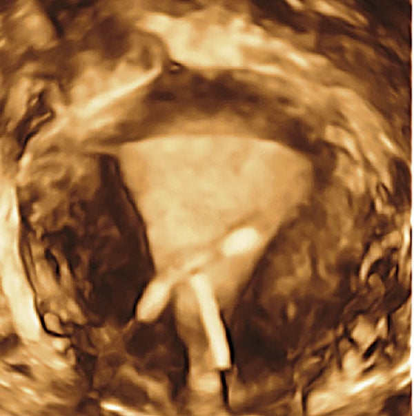
Coronal view, reconstructed from 3-D ultrasound, reveals an incorrectly positioned IUD. Image courtesy of American Journal of Obstetrics and Gynecology
That ultrasound offers benefits in cost, availability and minimizing radiation burden should come as no surprise. The medical community has been hearing this for a long time, especially during the past three years from the American Institute of Ultrasound in Medicine (AIUM) as part of its "Ultrasound First" campaign. What is surprising is that a group of opinion leaders in OB/GYN felt the need to advocate in the American Journal of Obstetrics and Gynecology (AJOG) the use of "ultrasound first" - specifically to image the female pelvis.
AIUM membership includes many of those who prescribe OB/GYN imaging studies in the United States (56 percent of its 10,000 members indicate an interest in obstetric ultrasound; 37 percent indicate an interest in gynecologic ultrasound). You might expect, therefore, that AIUM would be preaching to the choir when it comes to the cost-effectiveness and safety of ultrasound, particularly in regard to the female pelvis. Yet Beryl R. Benacerraf, M.D., and a half-dozen similarly respected colleagues found it necessary to state in the April issue of AJOG that "a skillfully performed and well-interpreted ultrasound image usually obviates the need to proceed to additional, more costly and complex cross-sectional imaging techniques," a tenet that applies "particularly to obstetric and gynecologic patients."
The need to state what might be considered obvious by many is palpable. As astounding as it may seem, computed tomography (CT) is the first choice for many women presenting with pelvic pain, masses or flank pain, the authors state in the AJOG article, just as magnetic resonance imaging (MRI) is the go-to modality for many with Mí¼llerian duct anomalies. Most distressing is that CT and MRI of the pelvis often yield indeterminate and confusing findings that require further clarification with ultrasound, the authors wrote.
"Doing a CT scan first for female patients with lower abdominal pain is dangerous, wasteful and expensive," said Benacerraf in a prepared statement. The clinical professor in obstetrics, gynecology, reproductive biology and radiology at Harvard Medical School and Brigham and Women's Hospital, went on to say, "We must educate the medical community to consider adopting 3-D ultrasound as the first assessment tool for specific gynecologic indications, such as evaluating the uterus for Mí¼llerian anomalies or localization of IUDs or other intracavitary lesions."
A decade ago 3-D ultrasound was more clinical curiosity than tool, limited by its dependence on the skill of the operator and restricted primarily to fetal assessment. Since then, however, volumetric scanning has matured. Its automated acquisition of operator-selected volumes can be sliced into hundreds of images reconstructed at just about any angle. Ultrasound is less expensive and less time-consuming than MRI, and free from the ionizing radiation of CT. And yet it provides many of the answers that these two modalities cannot.
A big part of the problem, according to AIUM Past-President Alfred Z. Abuhamad, M.D., is that many ultrasound practitioners have not yet embraced 3-D. Instead they continue providing basic 2-D scans. This "emphasizes the need for education and dissemination of this information," said Abuhamad, professor and chairman of obstetrics and gynecology at Eastern Virginia Medical School.
As America enters a new era of cost concerns, it is important that practitioners take advantage of tools that are effective clinically and financially, especially when those tools are best suited to answering their questions.
Greg Freiherr has reported on developments in radiology since 1983. He runs the consulting service, The Freiherr Group. Read more of his views on his blog at www.itnonline.com.

 June 24, 2026


