March 6, 2014 — Research shows that ultra-high-field magnetic resonance imaging (MRI) provides detailed views of a brain area implicated in Parkinson’s disease, possibly leading to earlier detection of the condition. The results of this research are published online in the journal Radiology.
With no radiologic techniques available to aid in diagnosis, clinicians have had to rely on medical history and neurological examination to detect Parkinson’s disease. It is often difficult to distinguish Parkinson’s disease from other conditions using these methods alone.
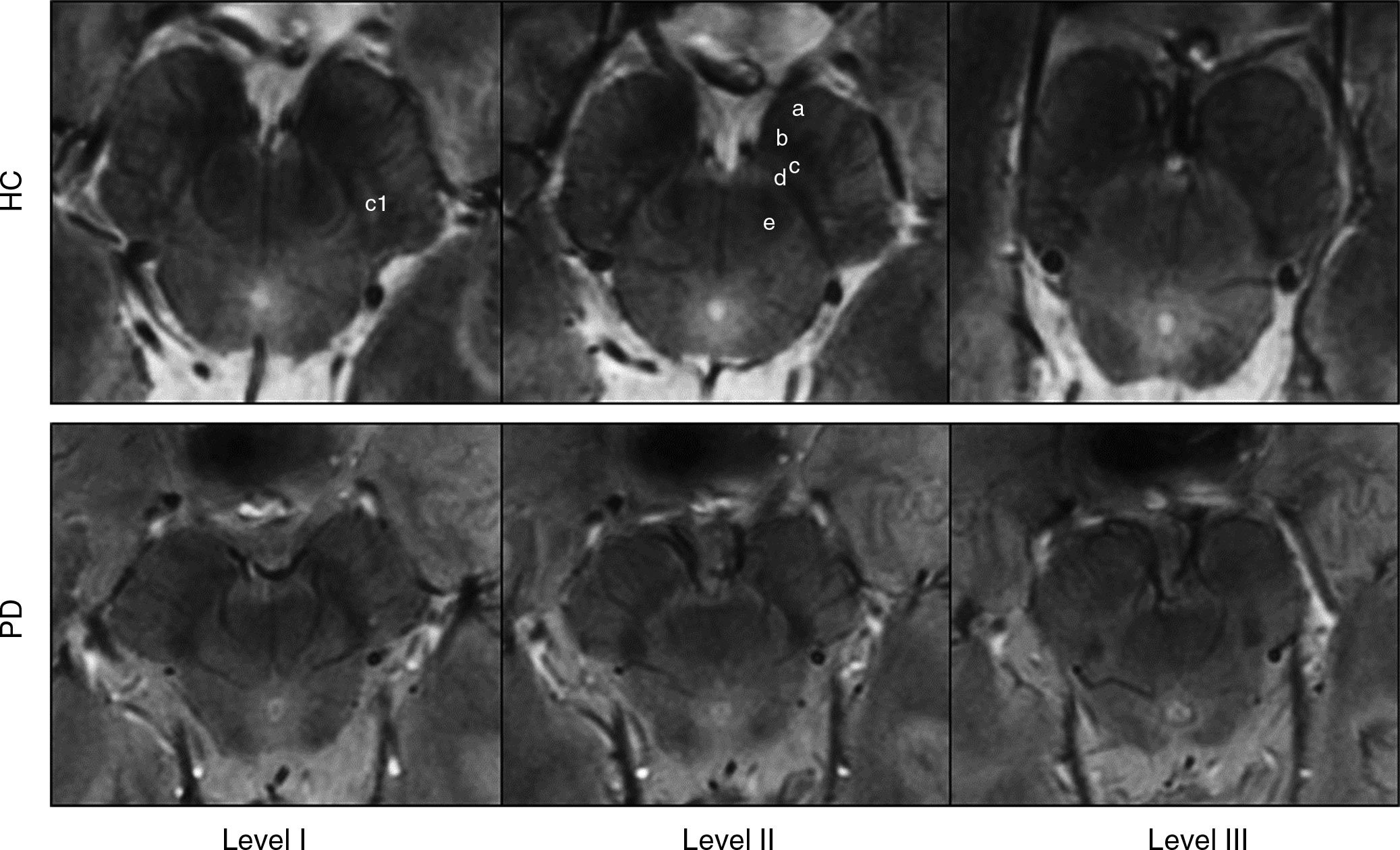
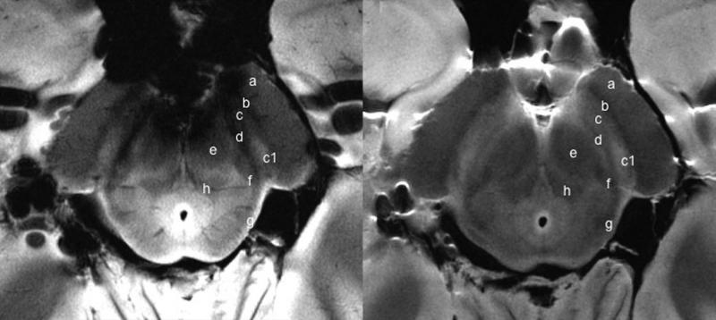
Mirco Cosottini, M.D., from the University of Pisa in Italy, and colleagues studied the brains of 38 individuals, including 17 Parkinson’s disease patients and 21 healthy controls, as well as a brain specimen from a deceased individual, to help determine the accuracy of ultra-high-field 7-Tesla (7-T) MRI for identifying Parkinson’s disease.
Using the 7-T MRI, the researchers were able to distinguish a three-layered organization of the substantia nigra (SN), a crescent-shaped mass of cells in the midbrain. Parkinson’s disease results from the loss of dopamine-producing cells located in this region of the brain.
Based on abnormalities in the SN identified by the 7-T MRI, the researchers correctly classified patients with Parkinson’s disease with a sensitivity of 100 percent and specificity of 96.2 percent.
According to Cosottini, the results show promise for earlier detection of the disease, which could speed the initiation of treatment.
“Parkinson’s disease diagnosis remains clinically based, but with the introduction of 7-T MRI into clinical practice, a supporting radiologic diagnosis can be made,” he said.
The researchers also are exploring the clinical utility of 7-T MRI in several other neurodegenerative diseases, including mild cognitive impairment, a precursor of Alzheimer’s disease.
For more information: RadiologyInfo.org

 June 12, 2026


