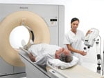
Photo courtesy of Philips Healthcare.
A report from the American Heart Association Committee on cardiac imaging stated that between 1980 and 2006 the collective dose from medical uses of radiation received by the U.S. population increased by more than 700 percent.1 The report also indicated “[Cardiac computed tomography angiography] (CCTA) accounted for around 50 percent of the collective dose.2
Moving forward, physicians need to introduce strategies that reduce the radiation dose patients receive, says professor Juhani Knuuti, the European Society of Cardiology’s working group on nuclear cardiology and CCTA.
Applying dose reduction strategies proved effective in the clinical trial PROTECTION 1.3 The study showed, for example, reducing the tube voltage from 120 kilovolts (kV) to 100 kV resulted in a 53 percent reduction in the median radiation dose for CCTA.
While using techniques to lower dose is an important first step, the biggest challenge is to standardize the use of radiation across hospitals throughout the United States.
To date, in the United States, there are no standard protocols for CT radiation dose exposure to patients during radiological procedures. Hospitals and imaging centers have developed their own protocols, typically based on those provided by their system’s manufacturer.
As a result, radiation dose varies from one facility to the next. One study found that although the estimated median radiation dose across many hospitals corresponded to 12mSv, there was a six-fold difference in the dose delivered between the highest and lowest centers.4
With growing awareness for the problem, is the medical community finally responding to the need for dose safety?
Growing Awareness
In the last 15 years, the problems with radiation exposure have escalated due to increased computed tomography (CT) utilization. A disproportionate amount of medical radiation dose comes from CT used in both pediatric and adult patients.
According to Carlos J. Sivit, M.D., vice chair for clinical operations, University Hospitals of Cleveland, Case Medical Center, part of the reason for the increased use of CT is because it is such a useful tool as a frontline test for patients of all ages.
After rising to its status as the study of choice in the mid-90s, by the early 2000’s, clinicians recognized there was an increase in radiation exposure to patients primarily driven by CT.
In February 2001, the American Journal of Radiology published a series of articles relating cancer risk to CT in pediatric patients, asking whether doctors were compensating for pediatric patients.
“This got the medical community, primarily the pediatric subset, and CT vendors to introduce certain interventions that could reduce dose,” said Sivit.
The public clamor eventually simmered down. However, in the last three years, there has been a steady resurgence in the debate. This time it has been driven as much by the pediatric side as by the adult side.
“It probably is as big of a problem in the adult population as it is in the pediatric population,” said Sivit. “We are most concerned with kids, since they have a greater time period to manifest results of radiation exposure. Children also tend to be more radiosensitive than adults. Because kids are so much smaller, using adult exposure parameters in pediatric exams results in significantly larger doses. This time around, there is heightened awareness in both the adult and pediatric imaging community.”
One of the thought leaders in the movement toward lower radiation dose is the Society for Pediatric Imaging (SPI). The society launched the ongoing Image Gently campaign to lower dose in pediatric patients. The campaign motto is “child-size” the amount of radiation used when obtaining a CT scan in children.5
Sivit believes Image Gently has had a significant impact, particularly on the vendors, the radiology community and on the public.
Yet, mainstream media continues to broadcast concerns with radiological procedures. The New York Times recently published articles revealing serious issues related to radiological procedures, in which radiation has harmed patients.
This prompted the House Energy and Commerce Committee’s subcommittee on health to hold a congressional hearing. Committee members examined incidents where excessive radiation doses were delivered during CT examinations and radiation therapy treatments. The committee noted that health care facilities across the nation lacked standards for technical personnel who perform medical radiation procedures, and needed uniform methods of reporting, investigating and tracking medical radiation errors.
“It’s totally unregulated. The sites are not inspected for parameters on how to use CT. They just want to make sure the seats are not dangerous. For the most part, they’re not, but they are capable of endangering people depending on how you use it,” he said.
The American College of Radiology proposed the development of a dose index to track the amount of dose a patient is exposed to over a lifetime. However, the challenge is how to measure dose.
Currently, there is not a direct method of measuring dose. There are two estimated values to measure dose. One is the CTDI (CT dose index), which measures absorbed dose to a specific tissue location. It allows comparison and evaluation of the effects of differences in technique relative to dose. There also is the dose length product, which is the dose index multiplied by the scan length. However, there is still no method to determine the cumulative dose that patients receive.
Strategies to Lowering Dose
Until radiation exposure in radiotherapy and medical imaging becomes standardized, there are several steps every doctor can take to safeguard patients.
Sivit provides six key strategies, from the beginning of the process to the end, that can diminish dose exposure.
1. Eliminate unnecessary exams:
The first step in dose reduction is to not use any dose by eliminating unnecessary exams or using alternative modalities that don’t use radiation. For example, apply ultrasound or magnetic resonance imaging (MRI). In addition, if there is a known condition, decrease the number of follow-up studies that use radiation whenever possible.
In many cases, probably 80 percent or more of the CT studies performed are normal or show incidental findings unrelated to the medical condition being considered. Decreased utilization is the best method of lowering dose.
2. Avoid using multiphase CT exams:
Whenever possible, avoid using multiphase CT exams.
Often, when doctors take a CT of the body, they may scan the patient two or three times. This may involve an arterial phase and portal venous phase exam. These protocols are associated with considerable dose. It is essentially getting three CTs instead of one, and they are rarely necessary. Multiphase exams are rarely used in the pediatric population, and could be eliminated in the adult population.
3. Adjust dose to patient size:
Modify the technique according to the patient’s size.
With CT, the higher the dose, the better the image will look. Radiologists, of course, want to have the best image possible, but it is not necessary to have the perfect image to make a diagnosis. This is widely acknowledged in the pediatric population, and many adult radiologists are catching on now.
The main driver of dose in CT is the tube current, which is the mAs. Optimize the tube current by keeping the mAs as low as possible.
Many systems provide automatic exposure control, which can reduce dose by as much as 50 percent. This feature forces the CT scanner to compensate for different parts of the body that may need less tube current. For dense anatomy, such as bone through the pelvis, the dose needs to be set at a higher tube current than when it is acquiring an image of the abdomen.
4. Calibrate exposure for key areas:
Change exposure to focus on key areas that answer the clinical question. For instance, when looking for lung or skeletal pathology, the user can lower the dose even more. In these areas, higher quality images don’t always lead to improved diagnosis.
5. Shield dose sensitive organs:
There are certain organs that are more dose sensitive: the eyes, the thyroid and the breast. Use external shielding to protect the breasts. For a facial exam, cover the eyes. When scanning the neck, shield the thyroid. This technique helps reduce dose to that organ, but doesn’t diminish image quality.
6. Pulse fluoroscopy and capture screen save images:
With fluoroscopy, the vendors have also made advances in the last decade. Instead of continuous fluoroscopy, use pulse fluoroscopy. The pulses can vary from 30 to three-and-a-half frames per second. Keep in mind: the lower the pulse, the lower the dose. That can decrease the effective dose by five- to 10-fold.
Instead of taking a spot image, use a screen saver. The screen saver captures the last object imaged and stays on the screen. By capturing an image of your screen saver, the patient is not exposed to any additional radiation.
While fluoroscopy does not generate as much radiation as CT, by applying pulsing or the screen saver technique, radiation levels are cut significantly.
Runaway Train
Meanwhile, CT utilization is increasing faster than the industry can regulate it.
“While there is greater awareness among radiologists and participation on the part of vendors, it’s almost like a runaway train that is hard to stop,” said Sivit. “The biggest factor right now is that CT is such a good test for so many things that utilization continues to increase dramatically.”
With a child or adolescent patient that is slender, standard parameters do not apply. However, many physicians do not adjust for their smaller size, don’t use automated dose reduction, and end up giving the patient 10 to 15 times the required dose. In 20 to 30 years, potentially that patient could develop disease from the radiation exposure. Sivit considers this a medical error.
“The medical definition of a medical error is ‘failure of a planned action to be completed as intended or use of a wrong plan to achieve an aim,’” noted Sivit. He feels overexposure to radiation should be considered as much an error as if a doctor gave a patient the wrong prescription.
The next step toward standardizing dose and quality control will require more cooperation from the vendors.
“The vendors need to provide the dose measurement,” said Sivit. “After that, software developers need to link the data into picture archive and communication systems (PACS) and the electronic medical record (EMR). Once dose is standardized, this would enable clinicians to better report and measure total patient exposure across multiple modalities over time.”
References:
1. Gerber, T., Arai , A., Gomes , A., et al. “Ionizing Radiation in Cardiac Imaging.” Circulation. 2009, 119:1056-1065.
2. Ibid.
3. “PROTECTION I: Radiation dose from cardiac CTA varied according to system, other factors.” Journal of the American medical Association. 2009;301:500-507.
4. Hausleiter, J. “Estimated Radiation Dose Associated With Cardiac CT Angiograph.” Journal of the American medical Association. 2009; 301:500-507.
5. Goske, M., et al. “The Image Gently Campaign: Working Together to Change Practice.” American Journal of Roentgenology. 2008; 190:273-274


 February 16, 2026


