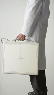
The Canon CXDI-50C Portable DR System is suitable for pediatric orthopedic use.
Imaging modalities and applications are going where they’ve never gone before. More than ever, healthcare providers are able to bring sophisticated imaging equipment to the patient, rather than vice versa.
Image quality, speed, flexibility of application and ease of use are highly valued by radiologists and other diagnostic clinicians, and the steady evolution of portable imaging systems leverages innovative technology and design to provide all these capabilities. Increased portability is congruent with the trend toward locating advanced technology at the point of care for easier, more accurate entry and retrieval of patient information. And portable imaging units usually complement, rather than replace, stationary imaging systems, while integrating with comprehensive information management processes.
Other drivers for portability include the convergence of technological advances that have brought us laptop computers and cell phones, along with market forces such as continual cost-control pressures and ever increasing patient expectations of high-quality healthcare. The move to miniaturization and digitization in electronics is continually balanced against the need for excellent quality and quantity of diagnostic data formerly thought impossible from machines this small.
The transition from film-based imaging equipment to digital systems usually enables faster patient throughput along with increased data storage capabilities and useful software applications, including image manipulation. State-of-the-art portable devices also deliver images that are higher-resolution than in the past, enable on-the-spot diagnoses and can dramatically shorten the time from evaluation to treatment in the emergency department, outpatient services and operating room.
Hospitals Move Toward Portability
Many hospitals and clinics are supplementing stationary modalities with portable equipment to improve their standard of care and reduce costs of in-hospital patient transportation, as well as process time, money and storage associated with conventional film systems. Some portable units are general-purpose, but most are designed for specific medical specialties.
The most popular applications for portable systems include emergency and sports medicine, obstetrics/gynecology, small clinics and remote locations, nursing homes and other situations where it is difficult for patients to be transported to stationary systems. The lightweight, modest environmental demands, flexibility and small footprint of portable units make them an attractive alternative for many clinical needs.
Each iteration of portable imaging systems, with its increased convenience, desirable features and improved clinical performance, is creating a broader range of applications and modalities, which is producing an increase in procedure volumes, including biopsy and surgery. These factors lead to an increased return on investment (ROI) and lower total cost of ownership (TCO) for portable units.
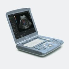
Ultrasound PCs Fit Multiple Environments
Ultrasound is critical to expediting patient throughput across the U.S. For hospitals with 200-plus beds, patient exams grew 41 percent from 13.1 million in 1998 to 18.5 million in 2005 — an average growth rate of five percent per year, according to a study by IMV Medical Information Division.
The importance of ultrasound in triaging emergency patients is growing stronger. “Our portable ultrasound is used for central intravenous line placement, thoracentesis, paracentesis and peripheral arterial pulse location,” said C.E. Ballard, M.D., an ED physician at Baptist Hospital in Little Rock, AR.
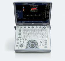
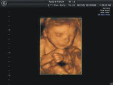
GE Healthcare is responding to the demand for ultrasound with plans to double its compact ultrasound business with the recent introduction of a new series of portable clinically specialized systems that provide sophisticated, real-time imaging at the point of care. Vivid i and Vivid e are designed for cardiology; Voluson i and Voluson E8 for obstetrics/gynecology; LOGIQ i and LOGIQ P5 have general radiology applications; and LOGIQ e is for the emergency room. The portable products pack powerful applications into lightweight, laptop-size designs with models like Voluson i that have 4-D visualization tools.
“Until now, the broad adoption of compact ultrasound has been hindered by image quality limitations and the industry’s ‘one size fits all’ approach to compact system design,” said Omar Ishrak, president and CEO of GE Healthcare’s Clinical Systems division. “By working with physicians from a wide-range of medical specialties, we’ve learned that image quality, portability and clinical specialization are all essential to expanding ultrasound’s role in healthcare.
“We’ve developed our new Compact Series to address these needs and bring the benefits of ultrasound to virtually all clinicians and patients – creating a pathway for ultrasound to become as ubiquitous in patient care as the stethoscope is today,” Ishrak continued.
Another advantage of miniaturized, portable units is that they are easily transportable within a hospital, ambulance or helicopter.
“[Portable systems are] perfectly adequate for 95 percent of our general purpose examinations,” said Peter Cooperberg, M.D., professor of Radiology at the University of British Columbia and chairman of Radiology for 500-bed St. Paul’s Hospital. “However, I don’t think that any radiology department would buy one of these machines as its only ultrasound unit. It is not a replacement for a full size machine.
“In our hospital, technicians only do normal ultrasound scanning, while trained doctors or sonographers do the portable imaging,” Dr. Cooperberg said. “We use portable ultrasound for any type of abdominal application, including pregnancies that need to be done while the patient is awaiting delivery. But mostly we use it in the ICU or ER for straightforward procedures like looking for abscesses or for perforations of blood vessels.”
Intuitive ease of operation and increased diagnostic accuracy are two of the portables’ appeals, especially in emergency situations. Cardiology-targeted portables are designed to be used as a visual stethoscope for the heart, says David Liang, M.D., an assistant professor of Cardiology at Stanford University. Physical examinations of the heart using a conventional stethoscope are notoriously unreliable, yielding 20 to 25 percent accuracy. However, studies at Stanford and Duke University indicate that using portable ultrasound produces diagnostic accuracy of heart exams from 50 to 65 percent.
New York’s Montefiore Medical Center’s Non-Invasive Vascular Laboratory, which treats a large volume of patients, has installed two portable Zonare zone ultrasound systems — compact, portable units that claim peak performance at a lower cost than conventional systems.
“Unlike other portable systems, Zonare’s breakthrough technology retains all of the raw data from the ultrasound signal,” said a Zonare spokesperson. “Consequently, a clinician is able to interrogate a complete diagnostic set of information after the exam and more closely scrutinize critical vascular structures or potential pathology.” The ultrasound system can be located at the bedside, operating room or anywhere in the hospital.
Advanced Systems Get Wheels
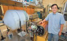
At the other end of the development spectrum, a laser-based MRI modality that would make the technology portable, quiet and cheaper has been developed by researchers with the U.S. Department of Energy’s Lawrence Berkeley National Laboratory in Berkeley, CA. The novel approach for MRI detection is based on optical atomic magnetometry. The technique provides a viable alternative for MRI detection with substantially enhanced signal-detection sensitivity at room temperature and time resolution for situations where traditional MRI is not optimal. Researchers hope that the laser-based MRI technique will make the technology compact, portable, relatively cheap and quiet.
Portable imaging systems are the main strategic focus of Ziehm, a C-Arm manufacture. Ziehm CEO Michael Calazoa said that larger vendors are developing portable products by just downsizing their stationary units, but that “Ziehm really has focused on the mobile aspects of this technology.”
Among Ziehm’s newer portable products are the Vision Flat, a fully digital mobile C-arm with a silicon-based flat-panel detector, and the new Ziehm Quantum. The latter is designed for surgical and interventional imaging applications, requiring fluoroscopy, including orthopedics, pain management, urology, neurology and vascular surgery.
Portable Imaging for Critical Care
On the treatment side, the Mobetron offers a fully portable, self-shielding device that directly delivers intraoperative electron radiation therapy to patients as they undergo surgery. Hospitals can wheel the Mobetron between operating rooms, eliminating the need for renovation that might be required by standard systems. Stanford Hospital and Clinics uses Mobetron for intraoperative electron radiation therapy that delivers high doses directly to a wide variety of cancers during surgery.
Breakaway Imaging has developed a patient-centric, O-arm imaging platform, designed for orthopedic and spine procedures, providing multimodality imaging: 2-D fluoroscopy, multiplane 2-D and 3-D at the patient’s side. The portable fluoroscope can be used in the operating room, ICU, emergency department, procedure rooms and trauma areas, with initial targeted applications of orthopedic and spinal surgical procedures.
New CRs and DRs Roll Out
Leveraging a rapid increase in digital technology to complement its film products, the Eastman Kodak Co. offers smaller DR and CR systems with a full range of applications, including dental and molecular. Kodak’s compact and flexible computed radiography (CR) products, designed for specialty markets such as orthopedics, diagnostic imaging centers and dentistry, are used for in-room needs throughout a medical facility, including emergency, pediatrics and intensive care departments.
At the same time, Kodak has introduced compact digital radiography (DR), such as the Direct View DR 7500, which touts its flexibility, versatility and economical cost.
“Emergency rooms today are overwhelmed with patients, so you have to handle more patients in less time. With an image every 20 seconds, you can see the workflow advantage of the DR,” said Phil Bullington, DR digital sales specialist for Kodak. “DR is faster and offers superior image quality. However, CR is less expensive and can service multiple X-ray rooms.”
Canon Medical Systems, a division of Canon U.S.A, will stage the introduction of its newest DR systems at RSNA 2006. The Canon CXDI-50C Portable DR System has a 14-by-17-inch image area with the LANMIT 7 (Large Area New-MIS Sensor and TFT) detector technology and is designed to deliver high-quality diagnostic images efficiently with minimal X-ray exposure to patients, making it suitable for pediatric orthopedic use.
Both of the ClearView CR readers used with Fuji’s CR for digital X-ray systems are used for mobile mammography imaging in Europe and Japan. The company expects to see them soon in the U.S. on FCRm mobile units, which received FDA approval for sale in July 2006. Additionally, Fuji’s DryPix 2000 and DryPix 4000 laser imagers were designed from the ground up for mobile imaging, especially mammography.
While the installed base of imaging modalities such as MR and PET continues to show healthy growth, portable imaging systems are becoming a valuable diagnostic adjunct and frequently utilized device both for clinical and cost considerations. Less processing time, data-rich applications and greater efficiency can result in better care and fewer errors while lowering costs for providers and patients.
Bill McGee, a 20-year healthcare industry veteran, has won national and regional awards for his writing on numerous medical topics.

 June 01, 2026