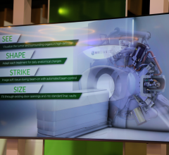
Editor’s Note: This is the result of a “virtual” roundtable discussion on image-guided radiation therapy (IGRT) with Drs. Rod Ellis, Dan Low, Todd McNutt, George Starkschall and Charles Smith. The first part appeared in the February/March 2006 edition of Outpatient Care Technology.
Please explain IGRT.
Ellis: IGRT is a moving target to see what’s in the literature. We started looking at image fusion to direct radiotherapy in 1997. We previously published our four- and five-year data, and presented our seven-year outcomes data at the ASTRO Translational Research Symposium in Radiation Oncology in San Francisco. It looks like IGRT is producing results that are better than standard radiotherapy. The technology is there, although it is still a challenge to work with some of the technology; there is a limitation in how the technology can be applied, but the time is now to start adapting to this technology.
Starkschall: A question that we must answer is what is our ultimate goal in using image-guided therapy? Just being able to look at the tumor prior to treatment and make some corrections for set-up uncertainty and tumor motion is not, in and of itself, an end — it’s a means to an end.
We would like to be able to increase the dose to the tumor or even better, control more cancer as a consequence of image guidance. Or conversely, decrease the dose that we are delivering to normal tissue and decrease the potential morbidity of the treatment. Until we can demonstrate either increased tumor control or decreased morbidity, we really haven’t proved the case for image-guided therapy.
Can you expand on the issue of portability
with studies?
Ellis: When I take a patient to the OR and perform an implant using ultrasound, I am trying to take data that was obtained in a supine position in radiology where they have been laid for a SPECT scan and a CAT scan, and performed image fusion. They take that information in an OR and put it in a dorsal lithotomy position, put a probe in the rectum, which may change the shape of the prostate slightly or by rotating the prostate slightly.
How do we account for those changes between the diagnostic image and the position that they are in for daily therapy, whether it be laying down on a radiation table for conformal radiotherapy or IMRT, or in the OR under ultrasound guidance? There has got to be a way to correlate and move these images from study to study and make the portability of these images between platforms.
Starkschall: One of the ways that people are trying to do this is through various kinds of imaging or image processing techniques such as deformable image registration. One of the problems with present day deformable image registration is some of the methods are based solely on the image. In applying such a technique, we make the assumption that just because a voxel has the same or similar number in one CT scan as in another CT scan, that the voxel represents the same chunk of tissue in both scans. Ultimately, we need to figure out a way to correlate the displacement of the actual tissue voxels rather than the CT voxels.
Do you have any thoughts or suggestions on ways to go about getting to that point — the ability to interpret the distortion of the tissue?
Starkschall: We’ve got some ideas. The deformable image registration algorithms may have to combine image information with perhaps deformable model approaches along with incorporating actual tissue properties into the deformation. Those techniques might help us get a better handle on how tissue voxels actually move.
It sounds like the hardware exists — we’ve got the imaging down — but it’s in the area of software that you don’t seem to be finding particularly innovative technology and would like to see more R & D at the vendor level. Is that correct?
McNutt: I think you are right. I think the tools are there, but what’s missing is the ability to manage the data in a way to make the tools efficient and reasonable in such a way that one can quickly analyze what they are seeing. It is an amazing amount of data. You are going from the potential of one CT scan of a patient for the whole course of treatment to 30 CT scans of that patient — the data explosion is huge. The ability to hold on to that data, analyze it and present it in a way that is efficient enough for a clinician to read quickly is where the big downfall lies.
To some extent even some computer technology may not be up to the challenge at this point. Some can argue radiology departments have huge PACS systems with tons of data and that’s okay for viewing images but it may be very difficult for doing full analysis of images.
Starkschall: We have a problem with this huge amount of data, and how to interpret this data. We may have 30 or more CT data sets for a given patient that need to be interpreted. How is the information we are acquiring going to be used to either adjust or not adjust the way we are treating the patient? All this information needs to be acquired and stored in a manner that can be readily retrieved as well.
Let’s explore the comment on developing in-vivo techniques and better contrast agents. Is there a belief that there is the ability to inject an in-vivo therapy into the body to attack the tumor and use the image to guide that treatment?
Ellis: What you are describing — injecting something to attack the tumors —that is exactly what I’ve been doing with brachytherapy by taking a monoclinic antibody and looking for a target. I think SPECT has been underutilized in oncology. I think PET will become better, but what other doctors and studies pointed out, is the absorption of the sugar is not necessarily anything specific to the tumor. With SPECT agents I believe you can have more specificity to a particular tumor.
So, for example, when you say you inject a monoclonal antibody, we use the image from the monoclonal antibody to see more clearly where the tumor is. Where I was heading with the in-vivo comment is to know whether the picture that you see with the functional image truly represents disease, and that if you had some markers that were placed, for example, at the site of a biopsy that shows known disease and areas of normal tissue, that may be a way to validate whether the functional study is giving a true volume.
What about developing better contrast agents?
Starkschall: In generating a treatment plan we have to deal with four major unknowns. The first of these is: Where is the tumor and what is the extent of the tumor? The other three are: What is the extent of the microscopic disease? How much does the tumor move during and between treatments? How much uncertainty is there in being able to set up the patient to treat the tumor? Let’s focus in on the tumor. Right now we are relying to a very large extent on the information we are getting from the CT scan to determine where the tumor is.
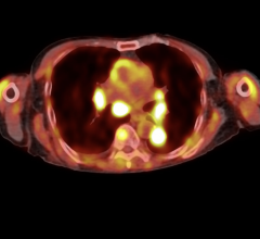
We’re using some of the other imaging modalities as a means to help us out, such as PET scans and SPECT scans. But the PET scan just tells you where the sugar is or where the sugar is being stabilized. It doesn’t really tell you unequivocally where the tumor cells are that are doing all the metabolizing. So somehow we have to determine some kind of contrast agent that will localize in the tumor and nowhere else, and be imaged by some type of imaging modality whether it is CT, PET, and SPECT. That is what I mean by better contrast agents.
Is it time to look into another way of delivering radiation therapy?
McNutt: I would argue a little differently that it’s not that we are delivering radiation therapy the same way we have for 15 years. Clinics have been spending all their time undertaking IMRT, which is by itself a total change in the way we deliver radiation therapy. So what is happening is just an evolutionary thing where currently the clinics are still finalizing how we’re doing IMRT and delivering IMRT and now there’s new technology, specifically image-guided therapy, and most of the clinics are still getting themselves fully engrossed in IMRT. That technology is exposing itself of the image guidance, and the early adopters are taking it on and now more clinics will start taking it on and will learn more and more and will learn better how to use it. But I wouldn’t make the argument that we have been doing things the same way for 15 years.
Can we take this data that we have today and use it to develop a future road map?
McNutt: Even then you have ultrasound image-guidance systems over the last decade and I think the use of electronic portal imaging devices have substantially increased since the emergence of the flat-panel detector. There are a lot of things that have occurred in image guidance. This new technology is more of an evolution than a revolution. We have used imaging for a long time. It has been evolving, we are just now getting to the point where we can see more about what we are doing.
Is IGRT the next logical step up from IMRT?
Starkschall: I wouldn’t look at it in terms of evolution; I think the two are running parallel. You can do IGRT with or without IMRT and you can perform IMRT with or without image guidance.
Smith: Initial and even current IMRT solutions present a clinically important paradox. It is possible to build a treatment plan that exceeds the ability to deliver it as precisely as it was planned if target position and/or motion isn’t properly accounted for. Like the emergence of CT imaging in treatment planning nearly a generation ago and IMRT in the last decade, IGRT has intuitive and dosimetric value.
Waiting for a complete solution to be derived for the IGRT “holy grail” may take years. If certain tools lead radiotherapy on the road to a global IGRT solution, they may add incremental value to patient care and would be worthy of consideration for implementation, once proven safe and effective. Using 4-D CT in treatment planning to more accurately characterize target motion during respiration and build treatment ports representative of that motion may be a good next step on that road. Of course, vendor collaboration with the academic community will go a long way toward ensuring that these tools are robust, cost-effective and ultimately usable in the mainstream clinical environment.
In terms of the development of techniques and technologies, what else do clinicians need to take IGRT to the next level?
Ellis: What we need is a way to get an image or report from radiology. You read the text and you know how to treat the patient. Now you are looking at 3-D volume. What we need to do is bring that 3-D volume to the treatment planning systems consistently and again, adapt for the fact that our patients are moving from day to day. Looking at one moment of a 3-D volume is not an adequate picture, whether it is anatomic or functional.
Low: I think automation is going to be key to adapting this technology. The matter of data, the complexity of moving it around and getting it between different systems is going to make it difficult for anybody with a lot of manpower and time and effort to implement on a wide basis.
Ellis: I think one last thing that is needed is a lot of continuing medical education and guidelines on how to apply this technology, so it gets into the clinic more rapidly.
From your viewpoint, what more can be done, what else needs to be explored? Is there a need for more intensive workshops throughout the year?
Ellis: I think years ago we used to all go to conferences and get our CME materials. Nowadays with the Internet we can sit down at our desk and download a video and get the CME. There are wider opportunities to get education materials out to the masses that are hungry for this knowledge.
Starkschall: It is very important for the radiation oncology community to recognize the need for training personnel in the implementation of these new technologies. Annually, I review applications for scholarships to the AAPM Summer School, which often addresses new technologies in either radiation oncology or in imaging.
Often I see on the application a letter from the administrator of a clinic saying they don’t have the money to train their physicists in the implementation of this new technology. The clinic is willing to spend a lot of money on the new technology but not willing to spend a few thousand dollars to make sure the individuals responsible for the safe and effective use of the new technology are adequately trained. I think we need to take a closer look at that.
Is there a concern that with this rapid expansion of technology, and the various levels of knowledge regarding technology, that we will come to a point where there will be great disparities? Is that something education needs to further address?
Smith: The education of physicians, physicists, dosimetrists and radiotherapists in both formal training programs and the broad clinical community will be critically important to successfully impacting the treatment of cancer with new imaging technologies in radiotherapy. A greater understanding of target motion will serve the medical staff in target delineation and dose prescription while clinical and technical integration of these systems and resources will fall largely on the physicist. Lastly, the treatment therapists will need to rely on an increased familiarity with cross-sectional imaging, image co-registration and other tools to effectively do their job. IGRT credentialing should deserve consideration and would help clinicians feel confident in their utilization or identify potential risks due to flawed methodology.
Any other thoughts on what we need to be focused on for the future of IGRT to take this technique to the next level?
Starkschall: We need to be very careful to make sure that we don’t introduce any unintended consequences as a result of incorporating IGRT into our clinical practice. We have image guidance to reduce margins down to 1 mm. However, in using image guidance to reduce our margins, we may have to repeat the imaging on a regular basis — maybe daily — to make sure that the treatment portal with the 1 mm margin is still valid. So we have to be careful how we use this imaging information in our clinical practice.
Feature | June 22, 2006 | rick dana barlow
Last of Two Parts
© Copyright Wainscot Media. All Rights Reserved.
Subscribe Now

 December 02, 2025

