With healthcare professionals thinking about the effects of ionizing radiation on the population at large, there is particular concern about its use for imaging children. That there is reason for concern was underscored by the release of new study results earlier this year, which indicated computed tomography (CT) exams of children in hospital emergency departments increased substantially from 1995 to 2008.[1] In addition, numerous studies have indicated ioninizing radiation has more serious effects on children than it does on adults, compounding the worry.
The industry has made concerted efforts to reduce radiation dose in pediatric imaging, especially from CT scans. These have included the establishment of the Image Gently campaign in 2007, an alliance for radiation safety in pediatric imaging supported by healthcare organizations around the world.
While CT still may be the best imaging choice in certain cases, there is continued emphasis on using other modalities whenever possible. As a result, magnetic resonance imaging (MRI) is being used more and more, according to Tariq Gill, M.D., chairman/medical director of diagnostic imaging, Lady of Lourdes Hospital in Binghamtom, N.Y., and president of Millennium Medical Imaging. “There is a new interest in using a modality that requires no radiation,” he said.
Besides the advantages of being radiation-free, MRI has other benefits for young patients, Dr. Gill noted. “With CT having the tunnel and the fact that parents can’t go into the room with the patient while the scan is being done, it can be more uncomfortable for the child,” he said. “With MR, parents can walk into the room and, especially with open MR, there is space so the child can see them during the scan. It makes a huge difference to the child in being able to go through the scan.
“MRI is becoming more and more the procedure of choice, whether it’s musculoskeletal or neurological, which are the most typical types of pediatric MRI exams,” he added. “The whole experience of scheduling these kids with less stress is improved.”
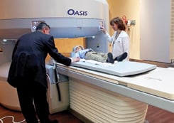
The open configuration where a child can keep visual contact with parents helps compensate for the fact that an MR exam takes longer than a CT scan. Lady of Lourdes Hospital has a Hitachi 1.2T open MRI system, and Dr. Gill said the open bore has been a tremendous help in creating a non-threatening environment. “It also has reduced our number of so-called failed studies, where we would end up having to sedate the child, which is always a concern,” he said.
“The fact that a caregiver can sit with the pediatric patient has meant that we have been able to scan them without sedation,” he added. “And the Hitachi system has a software algorithm that corrects for the little motion artifacts that might occur with a non-sedated child.”
Avoiding sedation not only is a benefit in terms of the pediatric patient’s well-being, but there also is a practical advantage. “If a child is uncooperative and we have to sedate him, that affects our throughput. It makes it hard,” Dr. Gill said. “In an outpatient setting, you can finagle the schedule around a little and accommodate the extra time that becomes necessary with sedation. But in a hospital setting, you never know what will happen. You could go from having no one waiting for a scan to having a dozen patients waiting all of a sudden. So throughput is an issue, and it is much improved and easier when there is no sedation required.”
Dr. Gill added that when they do have to sedate a pediatric patient, they look at the scans immediately to make sure they are good.
Technology Makes a Difference
The ability of MRI system manufacturers to develop an open configuration with high field strength has really changed things, according to Dr. Gill. “Previously, the magnets were low or intermediate strength,” he said. “With the high-field, with rare exceptions the open system images are comparable to the 1.5T closed systems now.
“With the 1.2T, we get a better signal-to-noise ratio, the scan is faster and slice thickness is thinner. So scan time is comparable to high-field 1.5T,” he added. “That’s a question we get asked a lot. It’s pretty much the same time.”
The ability to obtain good images from an open MRI not only is an advantage with pediatric cases; it also makes the imaging process more comfortable for bariatric and claustrophobic patients, as well as older patients who may be attached to life-support or other systems that must be accommodated.
“Theses four types of patients either fail the conventional mode of scanning or complete the exams without great success, with poor images,” Dr. Gill said. “With our new system, our MR volume has increased close to 50 percent because we are seeing those patients now.”
Easing the Burden for Parents
As Dr. Gill points out, pediatric cases also are unique because clinicians have to take parents into account as well as the patient. “You have to cater to the parents as well, even somewhat more than the actual patient,” he said. “When I had a child of my own, I really realized how a parent can be more nervous than the child.
“When parents are told their child might have a serious problem that requires testing, sometimes they end up traveling 80 or 100 miles to find a care center that can accommodate them. Even for people who are savvy travelers, going to a care center that’s far away from their home and is unfamiliar causes a lot of anxiety,” Dr. Gill added. “It adds stress to an already stressful situation.”
For him, having a system at Lady of Lourdes Hospital that can accommodate almost everyone in the community means “a huge burden has been lifted off our shoulders,” he said.
Reference
1 Larson DB, Johnson LW, Schnell BM, Goske MJ, Salisbury SR, Forman HP. “Rising Use of CT In Child Visits to the Emergency Department in the United States, 1995-2008.” Radiology. 2011 June; 259(3):793-801. Epub 2011 April 5.

 July 02, 2026


